Copyright
©2012 Baishideng Publishing Group Co.
World J Gastrointest Endosc. May 16, 2012; 4(5): 180-184
Published online May 16, 2012. doi: 10.4253/wjge.v4.i5.180
Published online May 16, 2012. doi: 10.4253/wjge.v4.i5.180
Figure 1 Endoscopic view of papillary large balloon (18 mm) dilation after limited sphincterotomy in a patient with a single large bile duct stone (30 mm, egg shaped).
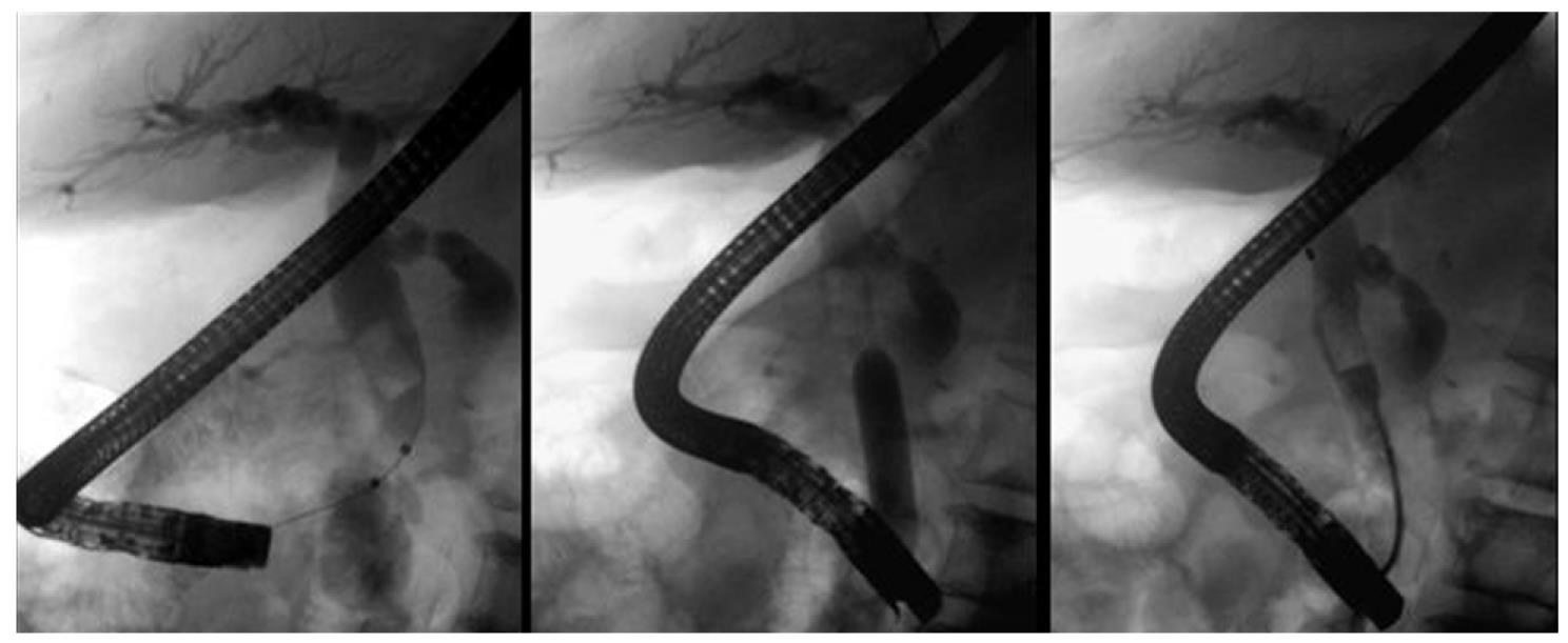
Figure 2 Fluoroscopy sequence showing a dilated common bile duct with a large single stone inside.
Balloon inflation until the notch on the waist disappears; clearance of the common bile duct with a Dormia basket.
- Citation: Rebelo A, Ribeiro PM, Correia AP, Cotter J. Endoscopic papillary large balloon dilation after limited sphincterotomy for difficult biliary stones. World J Gastrointest Endosc 2012; 4(5): 180-184
- URL: https://www.wjgnet.com/1948-5190/full/v4/i5/180.htm
- DOI: https://dx.doi.org/10.4253/wjge.v4.i5.180


