Copyright
©2012 Baishideng Publishing Group Co.
World J Gastrointest Endosc. Apr 16, 2012; 4(4): 151-156
Published online Apr 16, 2012. doi: 10.4253/wjge.v4.i4.151
Published online Apr 16, 2012. doi: 10.4253/wjge.v4.i4.151
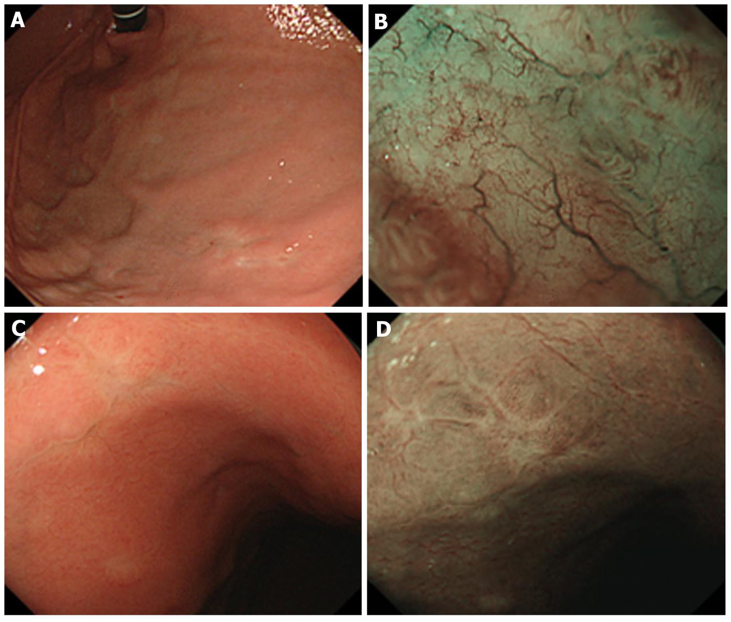
Figure 1 Routine observation revealed multiple ill-delineated brownish depressions in the stomach.
A: A brownish depression in the anterior wall of the upper gastric corpus; B: Narrow band imaging (NBI) findings of Figure 1A (TLA+); C: A brownish depression in the anterior wall of the middle portion of the gastric corpus; D: NBI findings of Figure 1C (TLA-). TLA: Tree-like appearance.
Figure 2 Routine observation revealed cobblestone-like mucosa at the greater curvature to the posterior wall of the upper gastric body.
A: Biopsy site of the cobblestone-like mucosa; B: Narrow band imaging (NBI) findings of the sites adjacent to the cobblestone-like mucosa at the upper gastric body (Figure 2A) TLA(-); C: Observation of cobblestone-like mucosa at the greater curvature to the posterior wall of the upper gastric body. Magnified endoscopy combined with NBI revealed disappearance of gland structure and branching abnormal blood vessels (Figure 2A) TLA(+). TLA: Tree-like appearance.
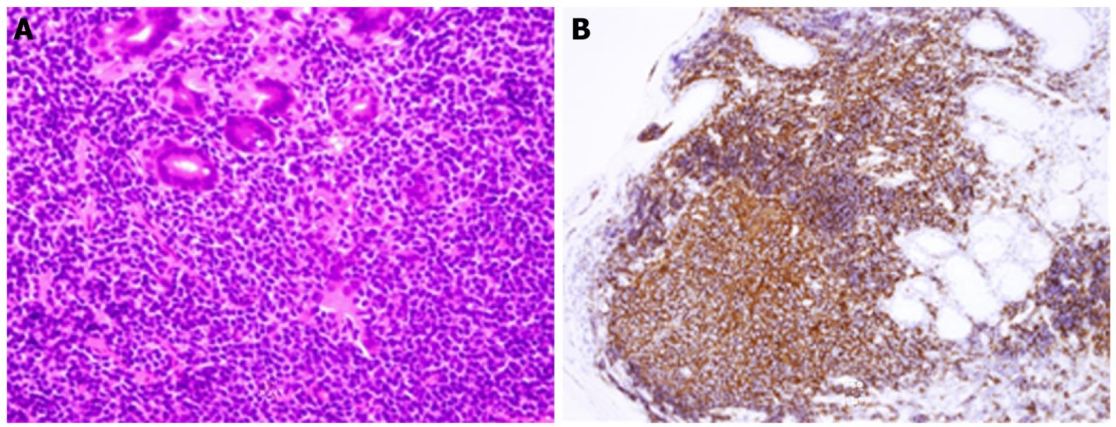
Figure 3 Histology of biopsy samples.
A: Histopathologic findings revealed hyperplasia of atypical centrocyte-like cells with small to medium-sized, ovoid nuclei and clear cytoplasm in the lamina propria mucosae and their infiltration among glandular epithelial cells, suggesting a lymphoepithelial lesion (hematoxylin and eosin stain, × 600); B: Immunohistochemistry. The lesion was positive for CD20 (× 200).
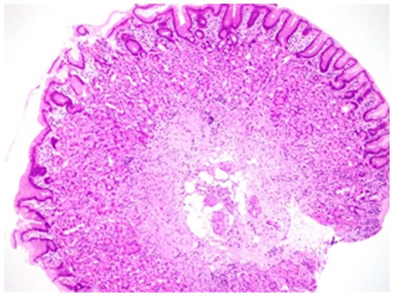
Figure 4 Histology of biopsy samples revealed only infiltration of inflammatory cells such as lymphocytes, plasma cells, and eosinophils in the gastric mucosa with foveolar hyperplasia (hematoxylin and eosin stain, ×100 ).
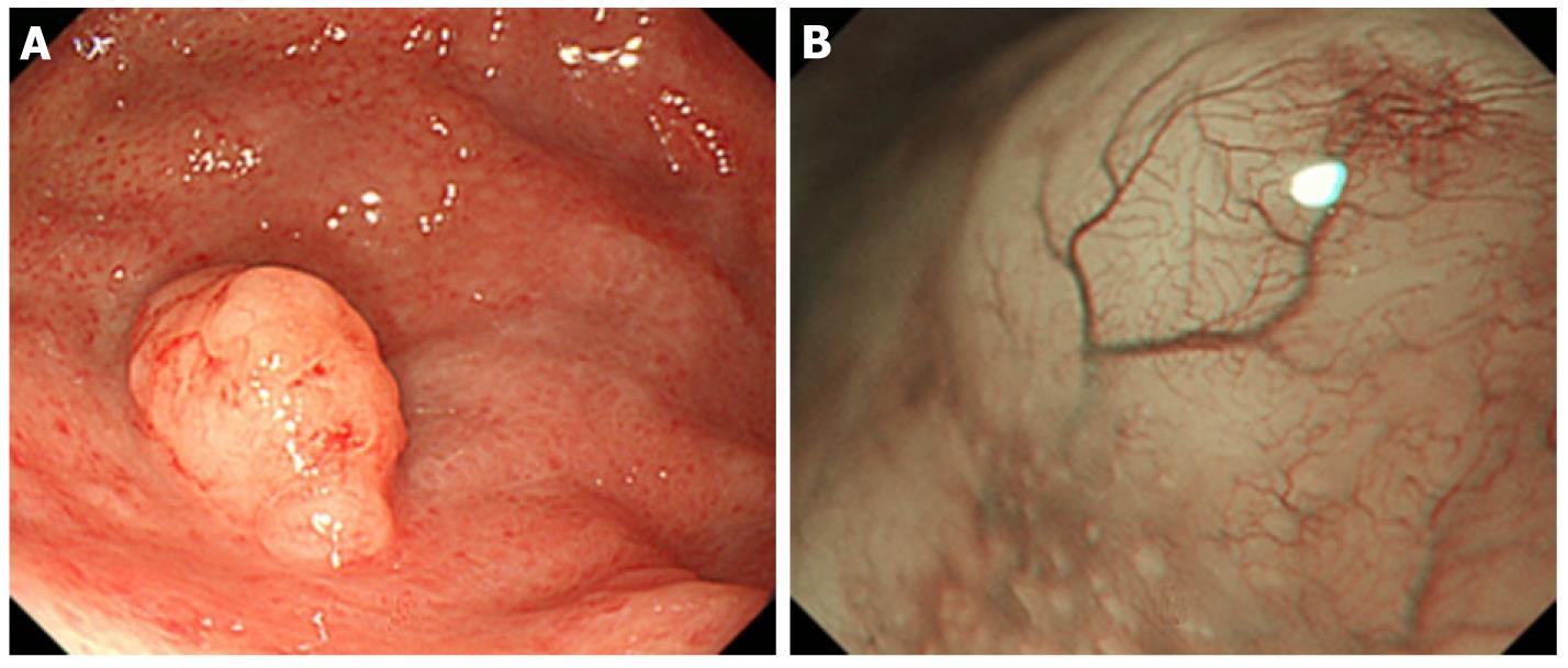
Figure 5 Endoscopic findings of gastric mucosa-associated lymphoid tissue lymphoma.
A: An elevated lesion, 10 mm in diameter, was observed in the greater curvature of the gastric fornix; B: Narrow band imaging findings. The mucosa that had lost its glandular structure, appeared shiny and had a tree-like appearance.
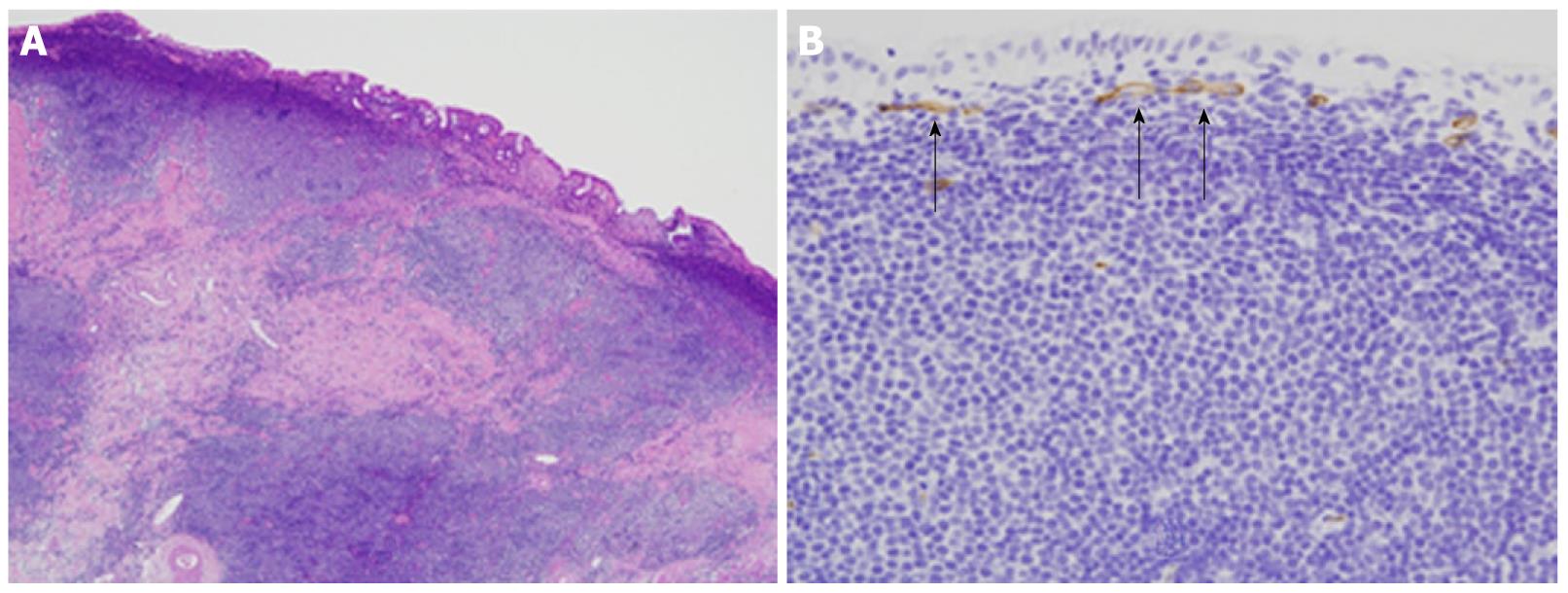
Figure 6 Histopathological examination of endoscopic mucosal resection specimen.
A: The mucosa was infiltrated by medium-sized atypical lymphocytes forming follicle-like structures. (hematoxylin and eosin stain, × 100 ); B: CD34. Unusual microvessels were observed running transversely immediately under the superficial layer of the mucosa (× 200).
- Citation: Nonaka K, Ishikawa K, Arai S, Nakao M, Shimizu M, Sakurai T, Nagata K, Nishimura M, Togawa O, Ochiai Y, Sasaki Y, Kita H. A case of gastric mucosa-associated lymphoid tissue lymphoma in which magnified endoscopy with narrow band imaging was useful in the diagnosis. World J Gastrointest Endosc 2012; 4(4): 151-156
- URL: https://www.wjgnet.com/1948-5190/full/v4/i4/151.htm
- DOI: https://dx.doi.org/10.4253/wjge.v4.i4.151


