Copyright
©2011 Baishideng Publishing Group Co.
World J Gastrointest Endosc. Jan 16, 2011; 3(1): 16-19
Published online Jan 16, 2011. doi: 10.4253/wjge.v3.i1.16
Published online Jan 16, 2011. doi: 10.4253/wjge.v3.i1.16
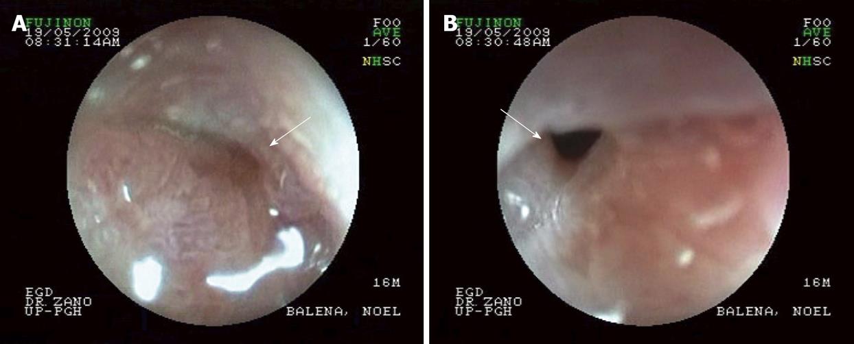
Figure 1 Endoscopic view of the 2nd part of the duodenal of the patient showing narrowing of lumen/stricture (white arrow).
The gastroscope was unable to pass beyond this point.
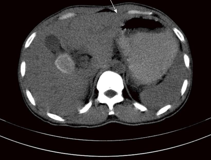
Figure 2 Abdominal computed tomography scan showing pyloroantral thickening and distended stomach.
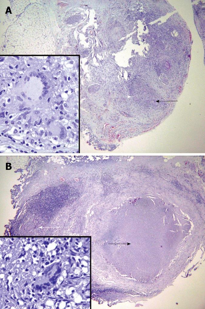
Figure 3 Histopathological examination image.
A: Duodenal mass showing chronic granulomatous inflammation (white arrow) with Langhans-type giant cell (black arrow); B: Calcified lymph node showing caseation necrosis (black arrow) and a multinucleated giant cell (inset, white arrow).
- Citation: Flores HB, Zano F, Ang EL, Estanislao N. Duodenal tuberculosis presenting as gastric outlet obstruction: A case report. World J Gastrointest Endosc 2011; 3(1): 16-19
- URL: https://www.wjgnet.com/1948-5190/full/v3/i1/16.htm
- DOI: https://dx.doi.org/10.4253/wjge.v3.i1.16


