Copyright
©2010 Baishideng Publishing Group Co.
World J Gastrointest Endosc. Sep 16, 2010; 2(9): 318-320
Published online Sep 16, 2010. doi: 10.4253/wjge.v2.i9.318
Published online Sep 16, 2010. doi: 10.4253/wjge.v2.i9.318
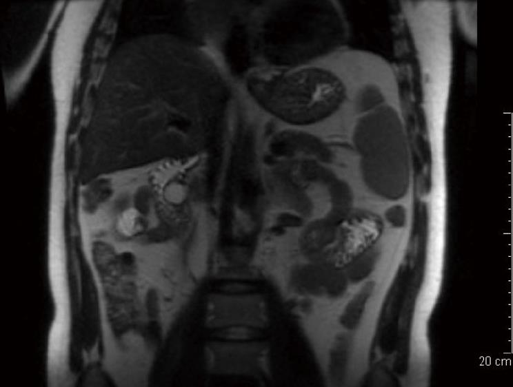
Figure 1 A cystic lesion in the area of the distal common bile duct, bulging into the duodenal lumen is seen in the magnetic resonance imaging.
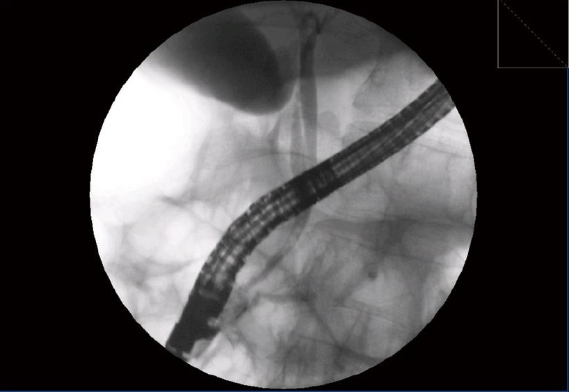
Figure 2 Pancreatography showed a blockage in the main pancreatic duct suggestive of pancreas divisum.
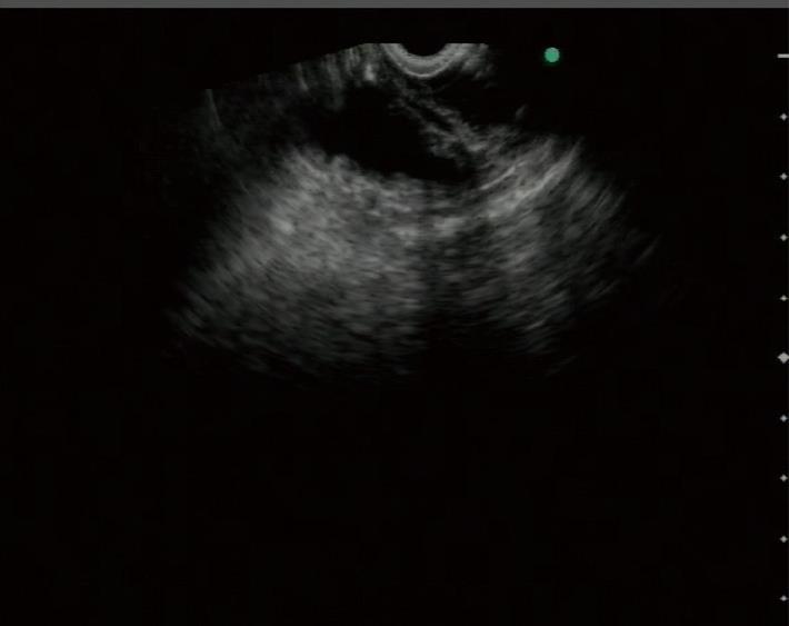
Figure 3 A cystic lesion found in the second duodenal portion, with three layer walls and debris inside the cystic cavity.
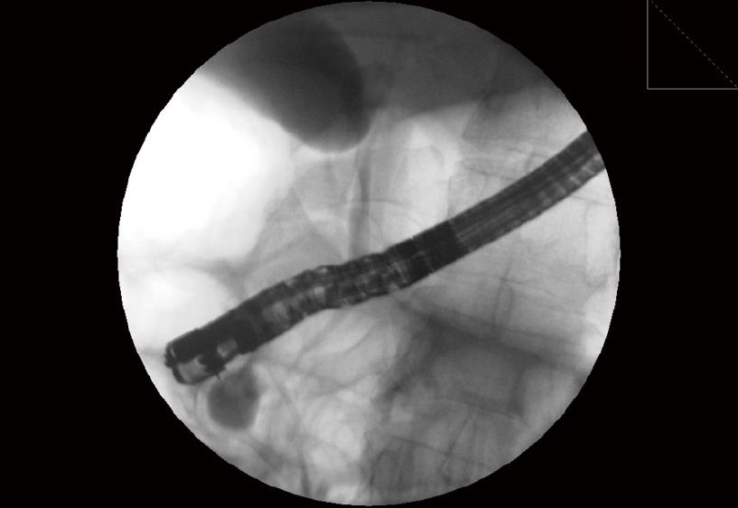
Figure 4 Contrast media is injected inside the cystic cavity with a needle knife.
- Citation: Redondo-Cerezo E, Pleguezuelo-Díaz J, Hierro ML, Macias-Sánchez JF, Ubiña CV, Martín-Rodríguez MDM, Teresa-Galván JD. Duodenal duplication cyst and pancreas divisum causing acute pancreatitis in an adult male. World J Gastrointest Endosc 2010; 2(9): 318-320
- URL: https://www.wjgnet.com/1948-5190/full/v2/i9/318.htm
- DOI: https://dx.doi.org/10.4253/wjge.v2.i9.318


