Copyright
©2010 Baishideng.
World J Gastrointest Endosc. Jun 16, 2010; 2(6): 223-227
Published online Jun 16, 2010. doi: 10.4253/wjge.v2.i6.223
Published online Jun 16, 2010. doi: 10.4253/wjge.v2.i6.223
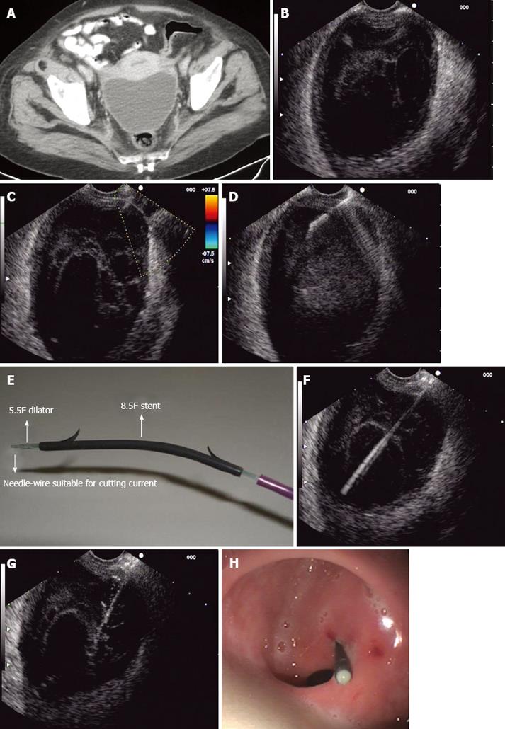
Figure 1 Endoscopic ultrasound (EUS)-guided pelvic abscess drai
nage procedure.
A: CT scan showing a pelvic collec tion at the Douglas pouch; B: Pelvic abscess detected by linear EUS; C: Color-Doppler showing no vessels between the abscess and the puncture site; D: Fineneedle aspiration with a 19-G needle; E: NWOA system for one-step drainage of fluid collections; F: 0.035-inch needle-wire into the abs
cess cavity (NWOA system); G: An 8.5-F stent inserted into the abscess (NWOA system); H: Successful drainage.
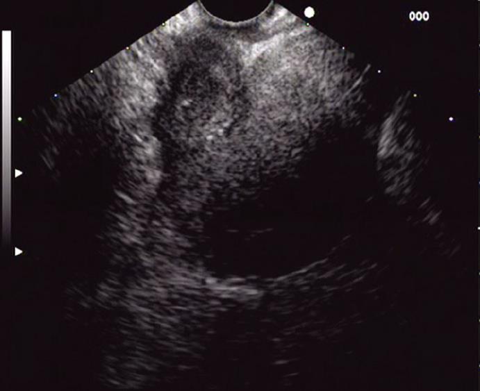
Figure 2 Virtual cavity close to the uterus after 3 wk.
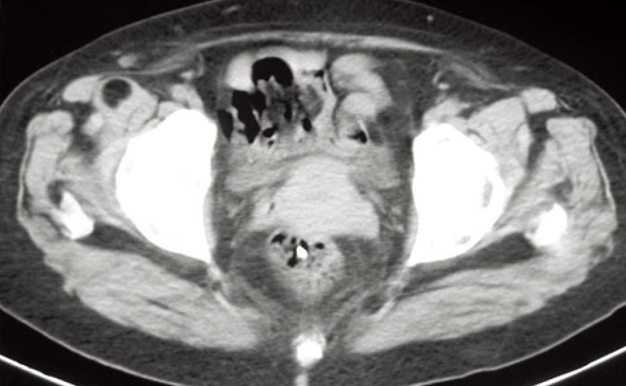
Figure 3 CT scan showing complete resolution of the abscess 4 wk later.
- Citation: Fernandez-Urien I, Vila JJ, Jimenez FJ. Endoscopic ultrasound-guided drainage of pelvic collections and abscesses. World J Gastrointest Endosc 2010; 2(6): 223-227
- URL: https://www.wjgnet.com/1948-5190/full/v2/i6/223.htm
- DOI: https://dx.doi.org/10.4253/wjge.v2.i6.223


