Copyright
©2010 Baishideng.
World J Gastrointest Endosc. Jun 16, 2010; 2(6): 198-202
Published online Jun 16, 2010. doi: 10.4253/wjge.v2.i6.198
Published online Jun 16, 2010. doi: 10.4253/wjge.v2.i6.198
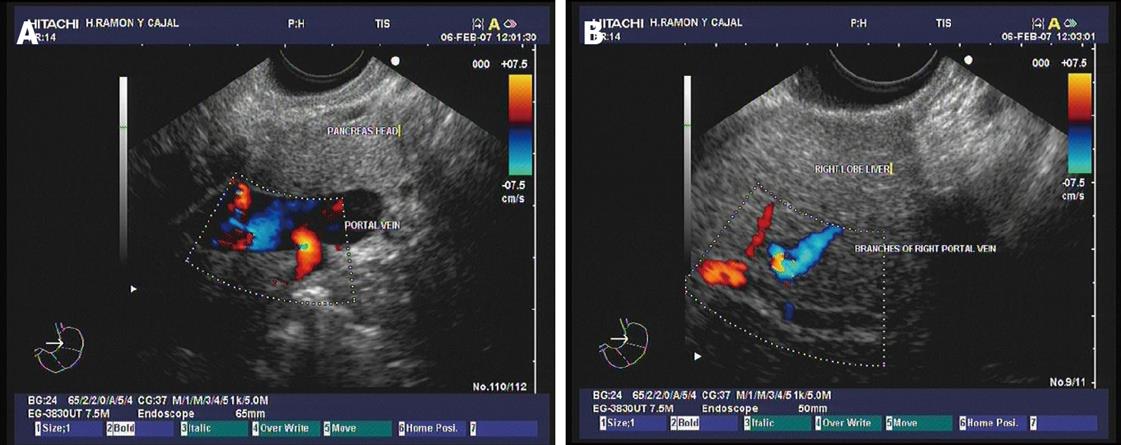
Figure 1 Doppler ultrasound images.
A: The main portal vein show adequate blood flow; B: Vein flow is also evidenced by interrogation of the branches of the right portal vein at the intrahepatic level. Images courtesy of Grupo Español de Protocolos en Endoscopia Digestiva.
Figure 2 Endoscopic ultrasound-guided embolization with coils of the right portal vein.
A: Image of the microcoil used in the experiment that is amenable for EUS-guided delivery[28]; B: The distal end of the echoendoscope is shown, showing the ultrasound transducer, the needle and the coil being deployed; C: EUS image of the right portal vein showing a grey coloured defect within the vessel that represents the coil after deployment; D: After coil deployment, X-ray fluoroscopy showed no filling of the right portal vein territory, with adequate contrast filling of the left portal vein. Images courtesy of Grupo Español de Protocolos en Endoscopia Digestiva.
- Citation: Vazquez-Sequeiros E, Olcina JRF. Endoscopic ultrasound guided vascular access and therapy: A promising indication. World J Gastrointest Endosc 2010; 2(6): 198-202
- URL: https://www.wjgnet.com/1948-5190/full/v2/i6/198.htm
- DOI: https://dx.doi.org/10.4253/wjge.v2.i6.198


