Copyright
©2010 Baishideng.
World J Gastrointest Endosc. May 16, 2010; 2(5): 190-192
Published online May 16, 2010. doi: 10.4253/wjge.v2.i5.190
Published online May 16, 2010. doi: 10.4253/wjge.v2.i5.190
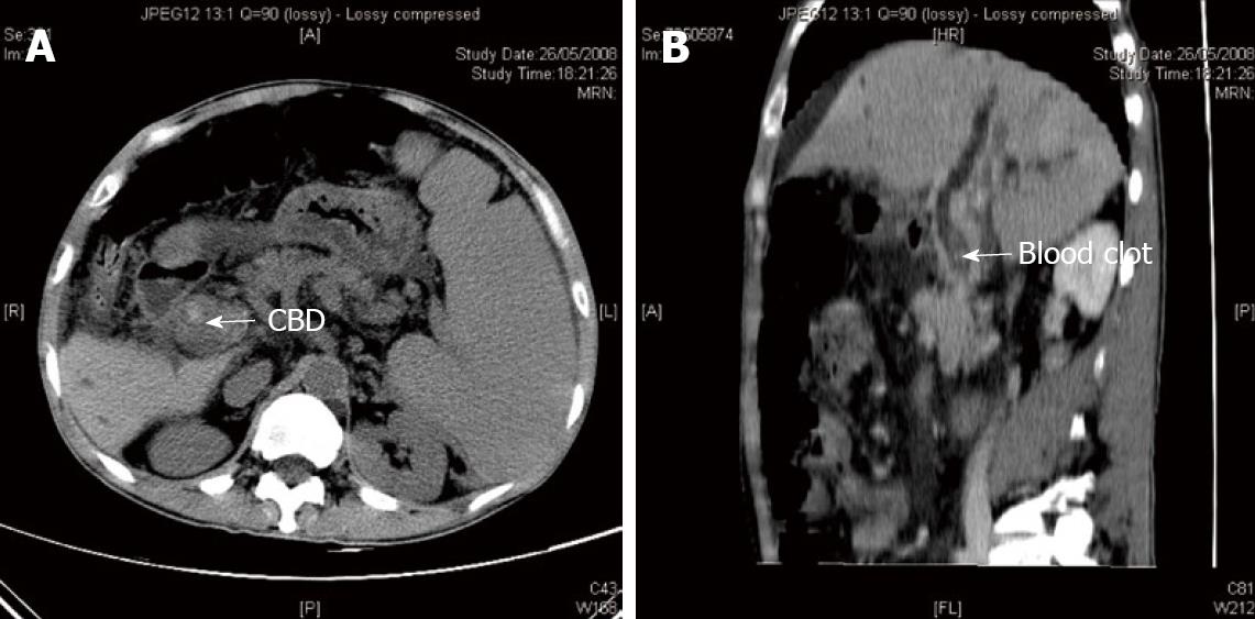
Figure 1 Abdominal computer tomography.
A: Non-contrast CT scan showing a hyperdense lesion inside the common bile duct causing upstream biliary dilatation; B: Contrast enhanced CT scan (sagittal view) showed a non-enhanced lesion inside the common bile duct.
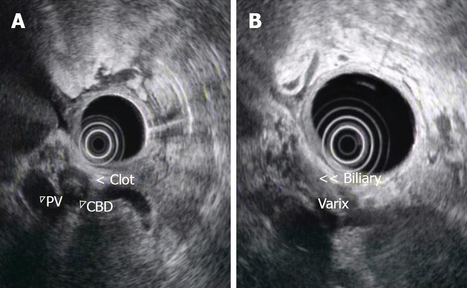
Figure 2 Endoscopic Ultrasound.
A: Endoscopic ultrasound image showing the CBD was completely obstructed by the hypoechoic clot; B: Endoscopic ultrasound image showing multiple small anechoic tubular structures around the CBD, consistent with varices.
- Citation: Ng CH, Lai L, Lok KH, Li KK, Szeto ML. Choledochal varices bleeding: A case report. World J Gastrointest Endosc 2010; 2(5): 190-192
- URL: https://www.wjgnet.com/1948-5190/full/v2/i5/190.htm
- DOI: https://dx.doi.org/10.4253/wjge.v2.i5.190


