Copyright
©2010 Baishideng.
World J Gastrointest Endosc. Mar 16, 2010; 2(3): 104-106
Published online Mar 16, 2010. doi: 10.4253/wjge.v2.i3.104
Published online Mar 16, 2010. doi: 10.4253/wjge.v2.i3.104
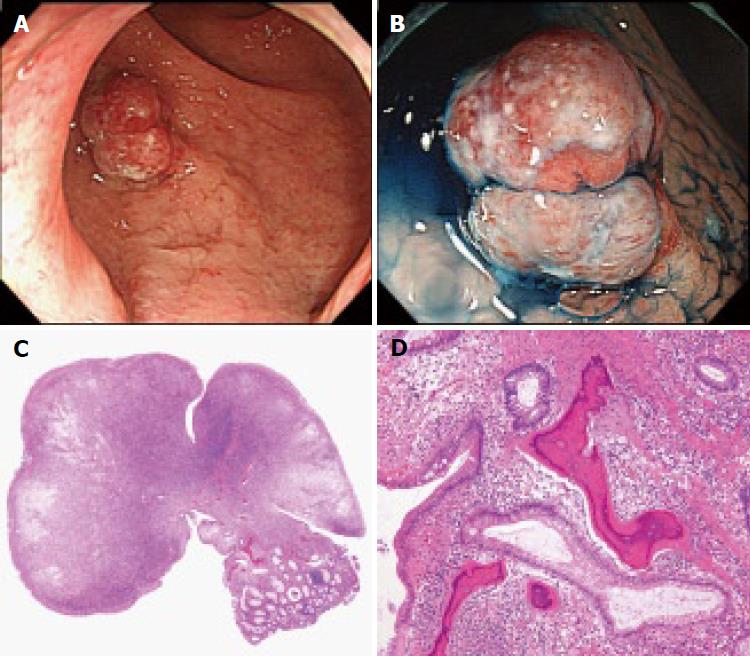
Figure 1 Subpedunculated polyp in the lower rectum.
A: Colonoscopy revealed a slightly reddish subpedunculated polyp, about 12 mm in diameter, in the lower rectum. The surface of the polyp was covered with whitish exudate, which suggested inflammatory change; B: Magnifying observation with dye-spraying using 0.4% indigo carmine revealed a type I pit pattern according to the Kudo’s classification; C: Histologically, the surface of the resected specimen was mostly covered by inflammatory exudate and partly by regenerating epithelium; D: Several foci of heterotopic bone formation were also found on histology.
- Citation: Oono Y, Fu KL, Nakamura H, Iriguchi Y, Oda J, Mizutani M, Yamamura A, Kishi D. Bone formation in a rectal inflammatory polyp. World J Gastrointest Endosc 2010; 2(3): 104-106
- URL: https://www.wjgnet.com/1948-5190/full/v2/i3/104.htm
- DOI: https://dx.doi.org/10.4253/wjge.v2.i3.104


