Copyright
©2010 Baishideng Publishing Group Co.
World J Gastrointest Endosc. Dec 16, 2010; 2(12): 413-416
Published online Dec 16, 2010. doi: 10.4253/wjge.v2.i12.413
Published online Dec 16, 2010. doi: 10.4253/wjge.v2.i12.413
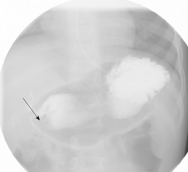
Figure 1 Filling defect of swollen pylorus (arrow).
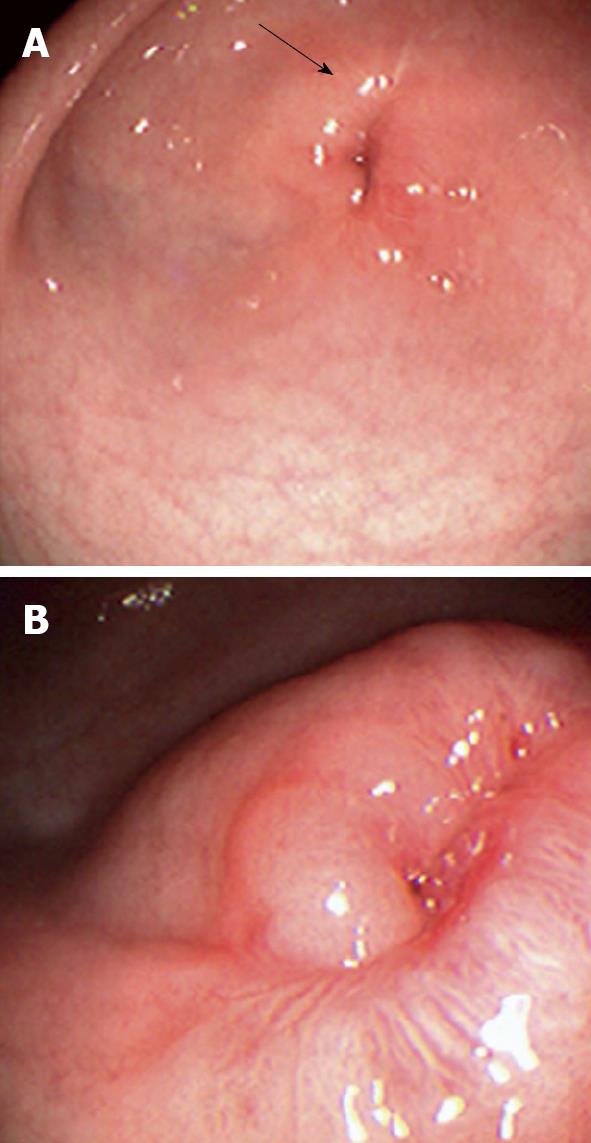
Figure 2 Upper gastrointestinal endoscopic image.
A: Narrow pyloric opening with edema around pyloric canal (arrow); B: Close-up view of pin-hole pyloric stenosis.
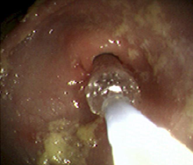
Figure 3 Pneumatic dilation across the pyloric channel.
-
Citation: Karnsakul W, Cannon ML, Gillespie S, Vaughan R. Idiopathic non-hypertrophic pyloric stenosis in an infant successfully treated
via endoscopic approach. World J Gastrointest Endosc 2010; 2(12): 413-416 - URL: https://www.wjgnet.com/1948-5190/full/v2/i12/413.htm
- DOI: https://dx.doi.org/10.4253/wjge.v2.i12.413


