Copyright
©2010 Baishideng Publishing Group Co.
World J Gastrointest Endosc. Dec 16, 2010; 2(12): 408-412
Published online Dec 16, 2010. doi: 10.4253/wjge.v2.i12.408
Published online Dec 16, 2010. doi: 10.4253/wjge.v2.i12.408
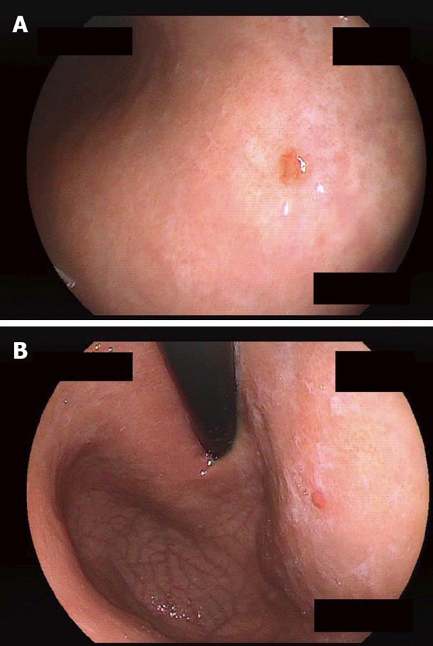
Figure 1 Tiny elevated lesions, 3 mm in diameter detected on the posterior (A) and the anterior (B) walls of the upper third of the stomach.
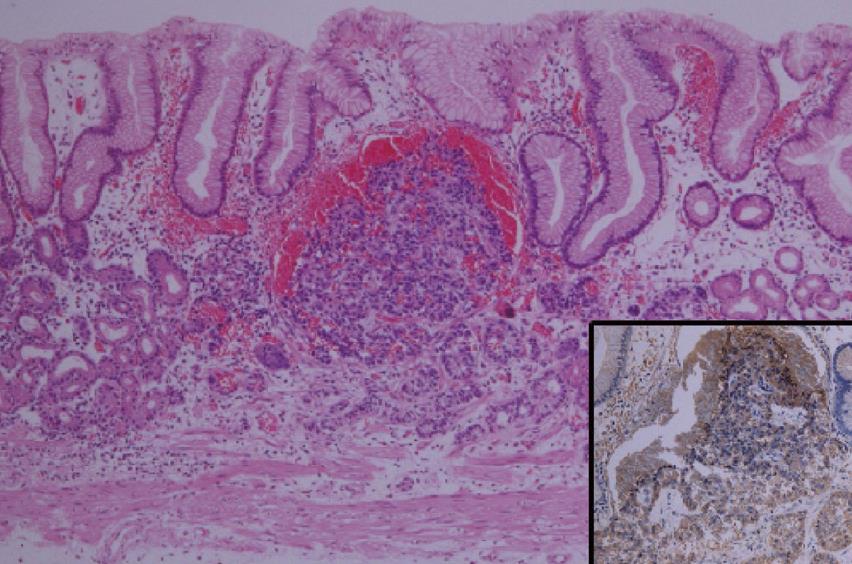
Figure 2 Histological findings of the tumor located on the posterior wall of the stomach (Hematoxylin-eosin stain, × 40).
The tumor exhibits microlobular-trabecular growth patterns with chromogranin A positive (inset, × 100). No cellular polymorphism is observed. The other tumor showed the same findings.
- Citation: Shimoyama S, Fujishiro M, Takazawa Y. Successful type-oriented endoscopic resection for gastric carcinoid tumors: A case report. World J Gastrointest Endosc 2010; 2(12): 408-412
- URL: https://www.wjgnet.com/1948-5190/full/v2/i12/408.htm
- DOI: https://dx.doi.org/10.4253/wjge.v2.i12.408


