
















Copyright: ©Author(s) 2026.
World J Gastrointest Endosc. Oct 16, 2026; 18(10): 122913
Published online Oct 16, 2026. doi: 10.4253/wjge.122913
Published online Oct 16, 2026. doi: 10.4253/wjge.122913
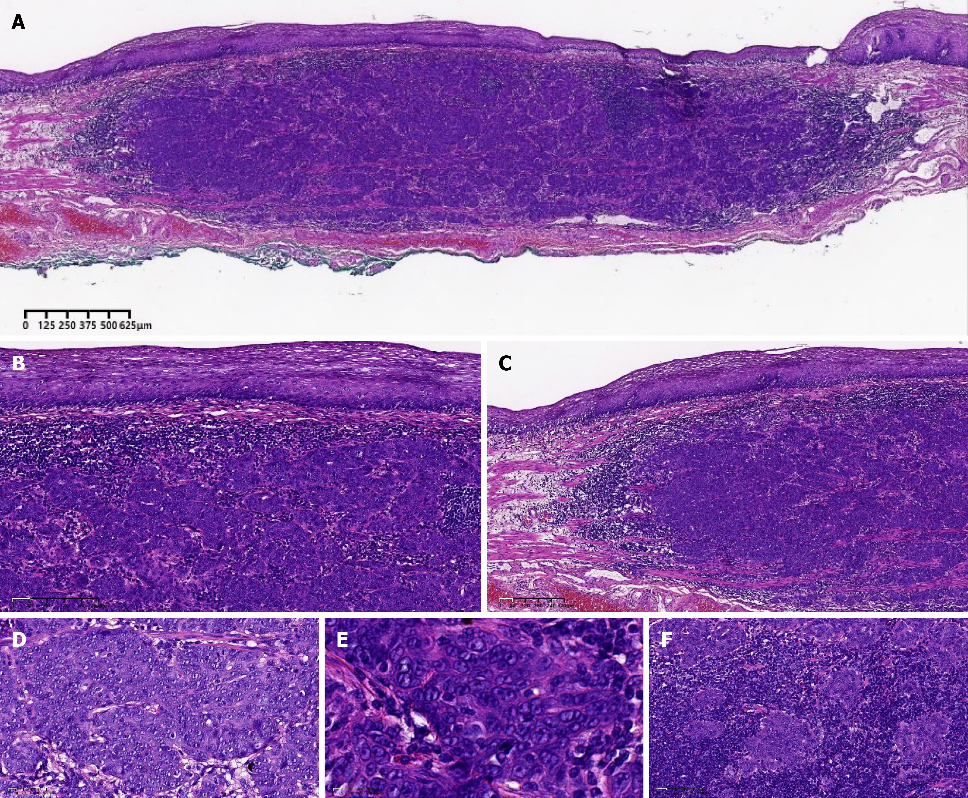
Figure 1 Pathology of the endoscopically resected specimen.
A: Beneath the mucosal epithelium, large areas of tumor parenchyma diffusely infiltrate in sheet-like and nested patterns; B and C: The tumor invades the lamina propria and muscularis mucosae, but the overlying epithelial cells are non-neoplastic; D and E: The tumor stroma is densely infiltrated by abundant mature lymphocytes and plasma cells, forming the characteristic architecture of “tumor nests embedded in abundant lymphoid stroma”; F: Tumor cells are large, with pale eosinophilic cytoplasm and ill-defined cell borders. The nuclei are round to oval, with thick and irregular nuclear membranes. Chromatin is pale and vacuolated, with prominent nucleoli; binucleated or multinucleated cells are observed in some areas. Mitotic figures are frequently seen, accompanied by marked cellular atypia. The white scale bars represent 40 μm, 60 μm, 10 μm, 5 μm, and 20 μm in B–F, respectively.
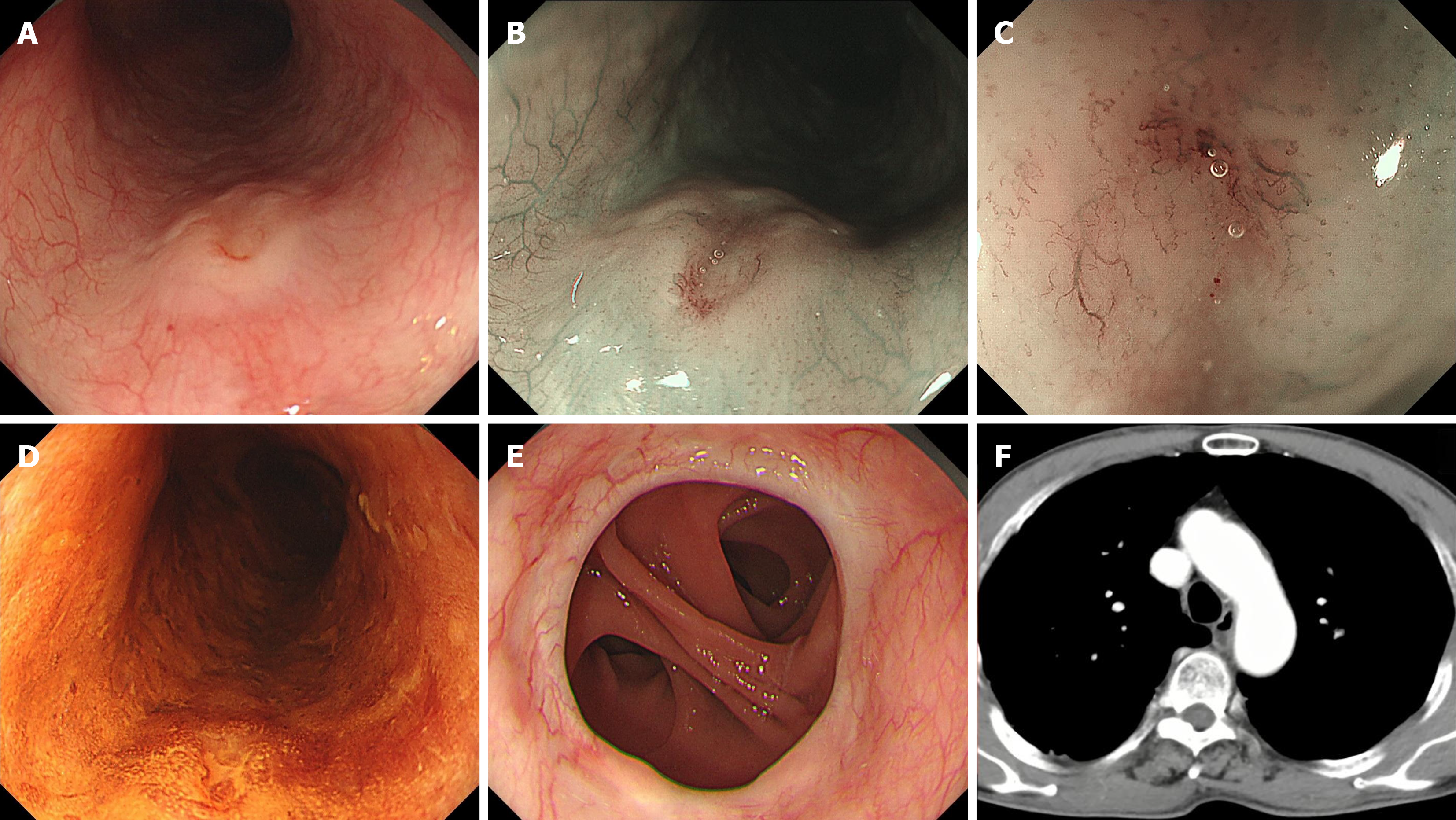
Figure 2 Endoscopic and radiographic findings before surgery.
A: Under white-light imaging, a pale type 0-IIa lesion is observed; B: Narrow-band imaging shows intact mucosa with no obvious demarcation, presumably indicating a submucosal origin; C: Magnifying endoscopy shows irregular B2i-like vessels; D: Lugol’s iodine staining shows a pale-stained area at the top of the lesion; E: Postoperative anatomy after total gastrectomy; F: Negative findings on computed tomography prompted the use of minimally invasive endoscopic submucosal dissection.
- Citation: Mou Y, Li S, Zhang YH, Hu B. Endoscopic submucosal dissection combined with radiotherapy for T1b esophageal lymphoepithelioma-like carcinoma: A case report. World J Gastrointest Endosc 2026; 18(10): 122913
- URL: https://www.wjgnet.com/1948-5190/full/v18/i10/122913.htm
- DOI: https://dx.doi.org/10.4253/wjge.122913