Copyright
©The Author(s) 2025.
World J Gastrointest Endosc. May 16, 2025; 17(5): 101618
Published online May 16, 2025. doi: 10.4253/wjge.v17.i5.101618
Published online May 16, 2025. doi: 10.4253/wjge.v17.i5.101618
Figure 1 Gastroscopy and colonoscopy.
A: There was no bile retention in the gastric cavity, the gastric mucosa was smooth; B: The mucosa of the descending part of the duodenum was rough; C: Colonoscopy showed smooth colorectal mucosa; D: Lymphoid follicular hyperplasia was seen at the end of the ileum, which was scattered and multifocal and relatively homogeneous in morphology.
Figure 2 Whole-small bowel double-balloon enteroscopy.
A: Roughness of the mucosa of the descending duodenum; B: Cracking changes of the villi after indigo carmine staining of the descending duodenum; C: Roughness and swelling of the mucosa of the horizontal segment of the duodenum, with cracking and mosaic changes of the villi atrophy; D: Mosaic-like changes in the jejunal mucosa.
Figure 3 Whole-small bowel double-balloon enteroscopy.
A: Multiple segmental longitudinal mucosal lesions in the ileal segment; B-D: Mucosal lesions after indigo carmine staining at a distance and close up: Some of the villi on the surface were detached, there was no white moss or exudation at the base, and the mucosal swelling at the edge of the base was obvious as a nodule-like bulge, and the ileal villi around the lesion were flattened and atrophied.
Figure 4 Whole-small bowel double-balloon enteroscopy.
A-D: Scattered multiple lymphoid follicular hyperplasia is seen in the ileum, with lymphoid follicles becoming progressively larger and denser from the proximal to the distal part.
Figure 5 HE staining of small intestinal mucosa.
A and B: The villous structure becomes blunt, the crypt structure extends, and the number of lymphocytes in the surface epithelium increases, but the number of plasma cells in the lamina propria decreases or is missing; C and D: Increased intraepithelial and intrinsic membrane lymphocytes, with some showing focal lymphocyte proliferation.
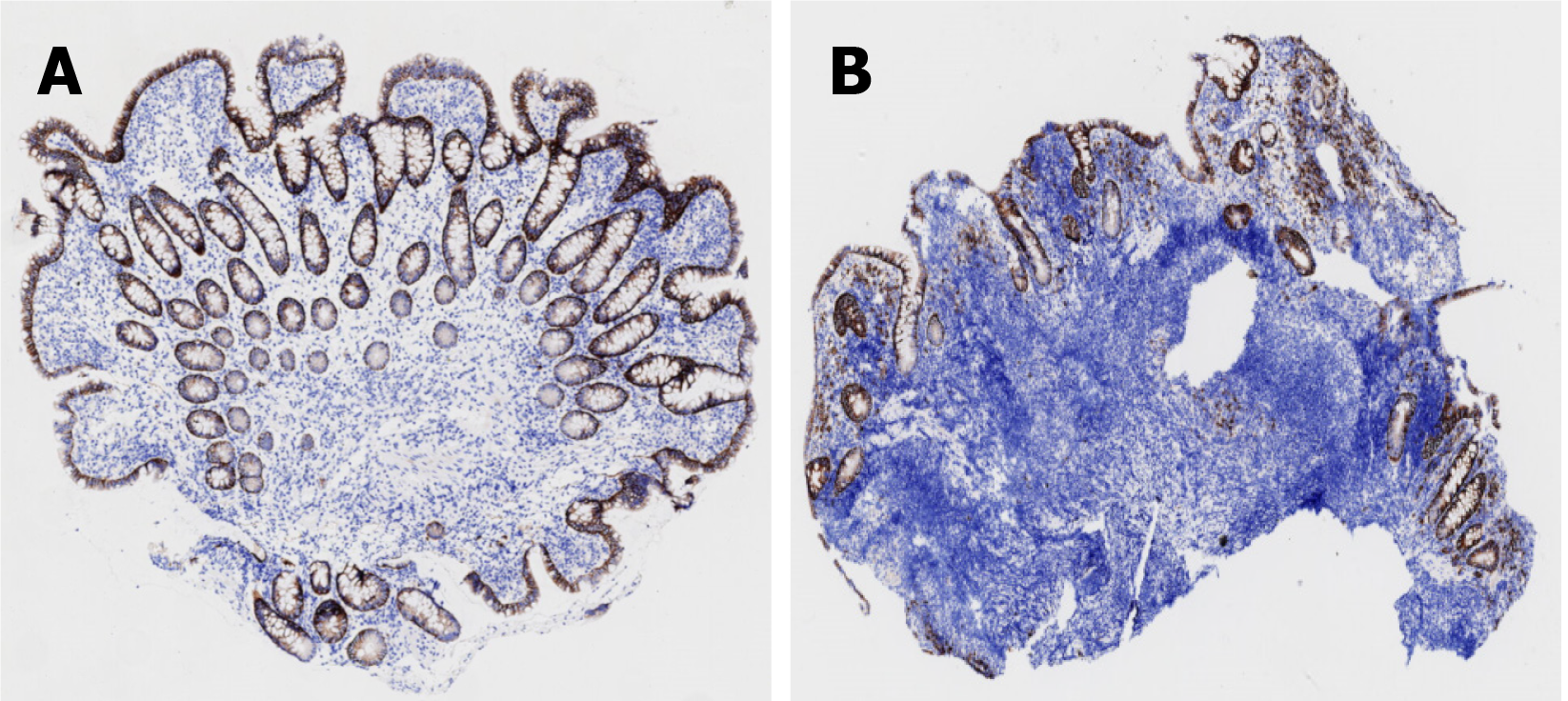
Figure 6 Immunohistochemistry.
A and B: CD138 staining was negative in lamina propria mucosa.
- Citation: He T, Fan MM, Zhang PQ, Zhang W, Fan D, Du LS, Tang M, Wan P, Song ZJ. Diverse phenotypic manifestations of small intestinal mucosa in non-infectious common variable immunodeficiency bowel disease: A case report. World J Gastrointest Endosc 2025; 17(5): 101618
- URL: https://www.wjgnet.com/1948-5190/full/v17/i5/101618.htm
- DOI: https://dx.doi.org/10.4253/wjge.v17.i5.101618


