Copyright
©The Author(s) 2024.
World J Gastrointest Endosc. Jan 16, 2024; 16(1): 44-50
Published online Jan 16, 2024. doi: 10.4253/wjge.v16.i1.44
Published online Jan 16, 2024. doi: 10.4253/wjge.v16.i1.44
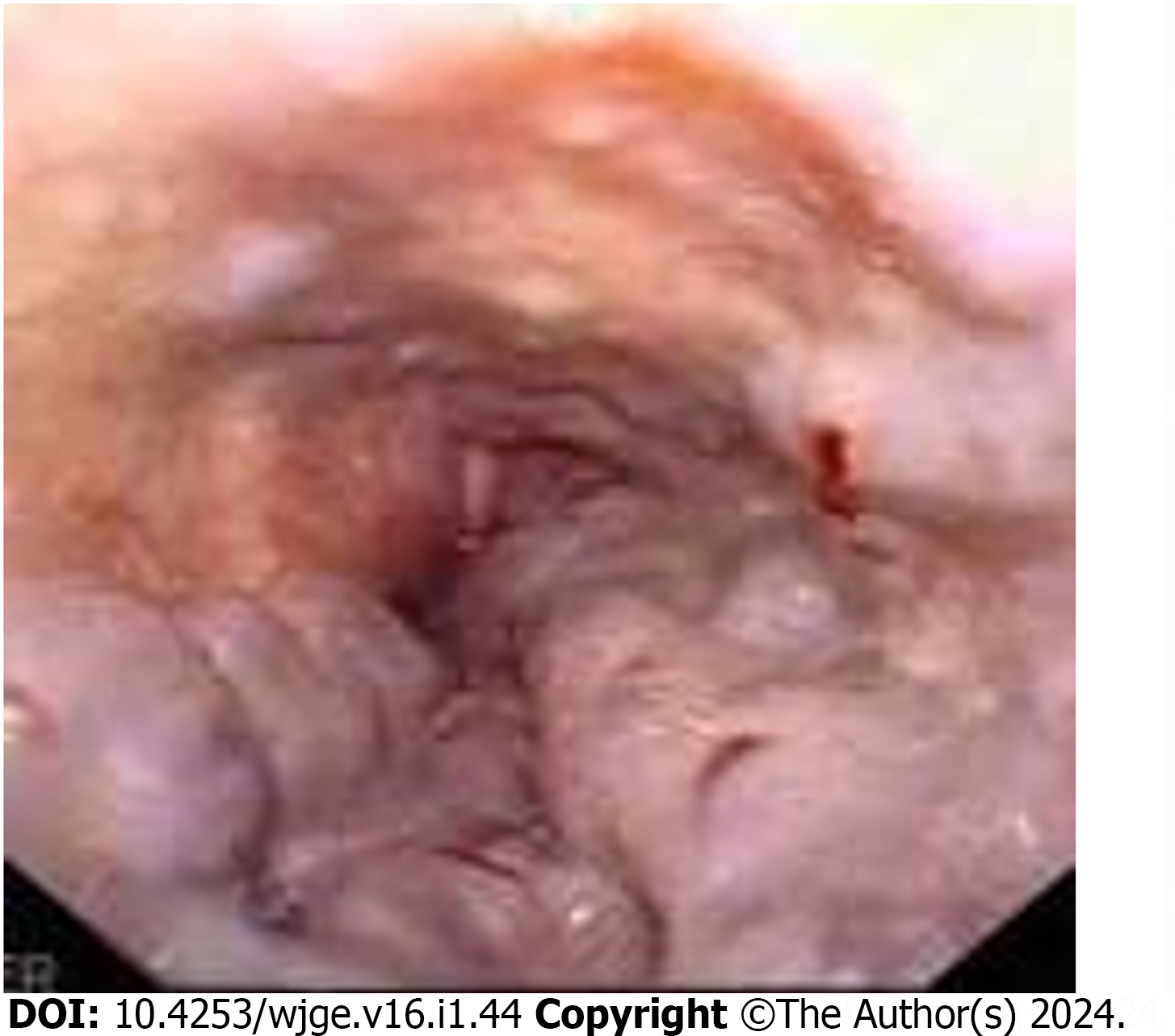
Figure 1
Example of grade 4 esophageal varices.
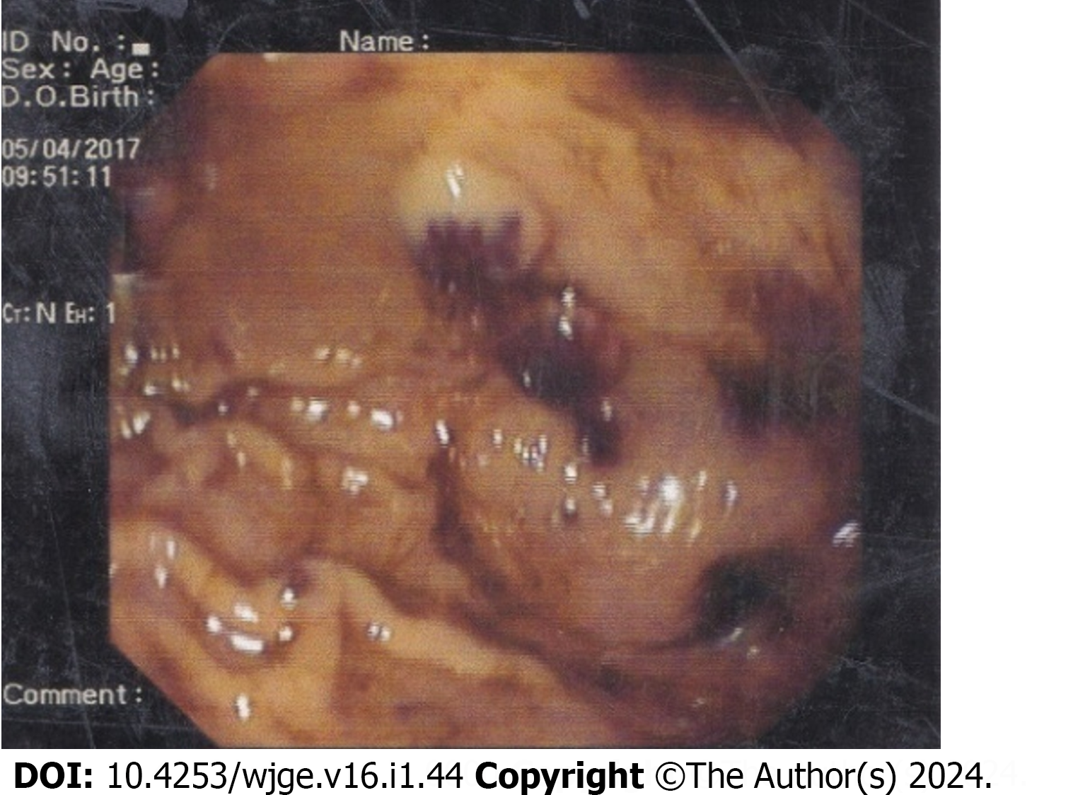
Figure 2
Blue rubber bleb nevus in the stomach.
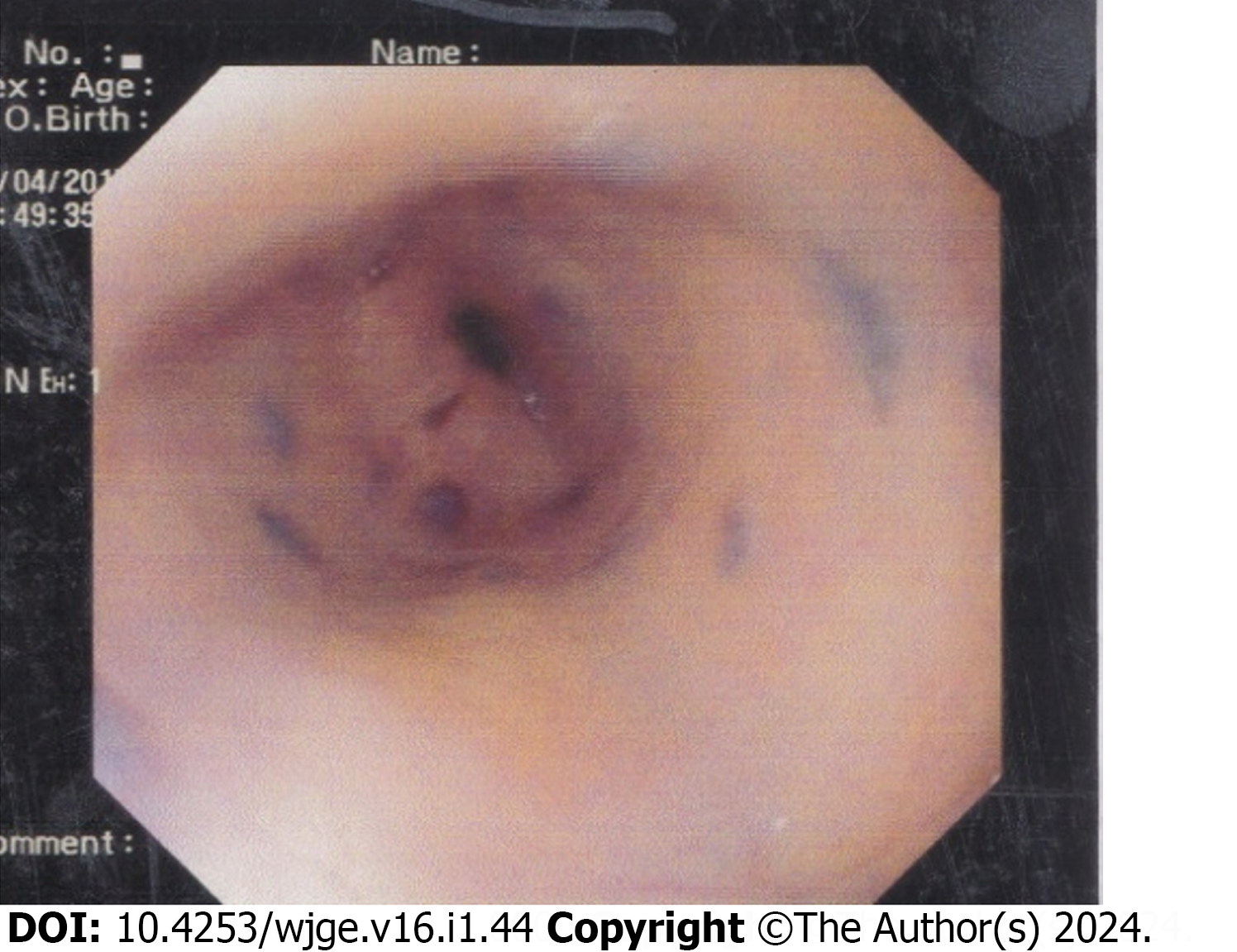
Figure 3
Blue rubber bleb nevus in the lower end of the esophagus.
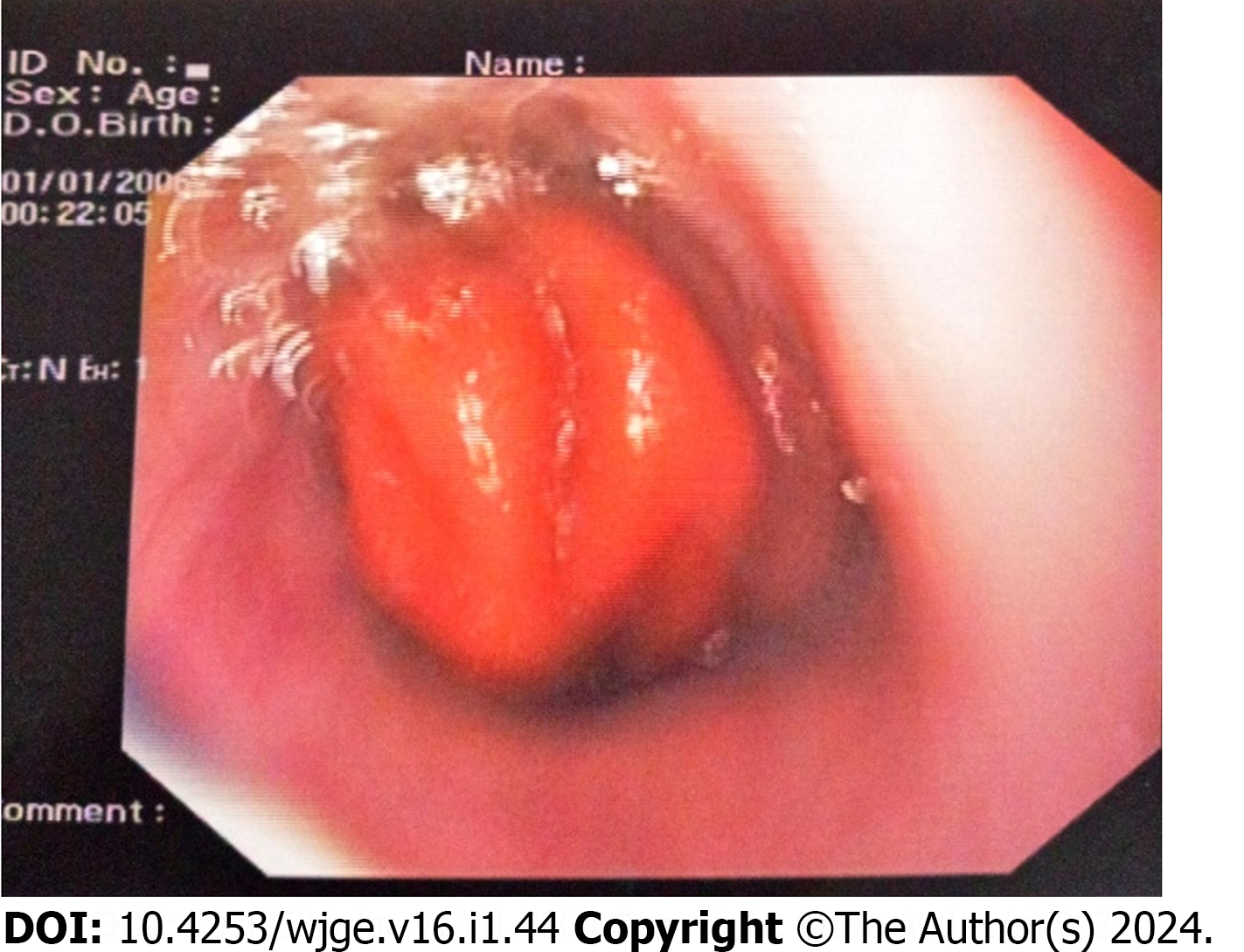
Figure 4
Prolapsed fundus of the stomach in the esophagus.
- Citation: Mazumder MW, Benzamin M. Upper gastrointestinal bleeding in Bangladeshi children: Analysis of 100 cases. World J Gastrointest Endosc 2024; 16(1): 44-50
- URL: https://www.wjgnet.com/1948-5190/full/v16/i1/44.htm
- DOI: https://dx.doi.org/10.4253/wjge.v16.i1.44


