Copyright
©The Author(s) 2023.
World J Gastrointest Endosc. Aug 16, 2023; 15(8): 540-544
Published online Aug 16, 2023. doi: 10.4253/wjge.v15.i8.540
Published online Aug 16, 2023. doi: 10.4253/wjge.v15.i8.540
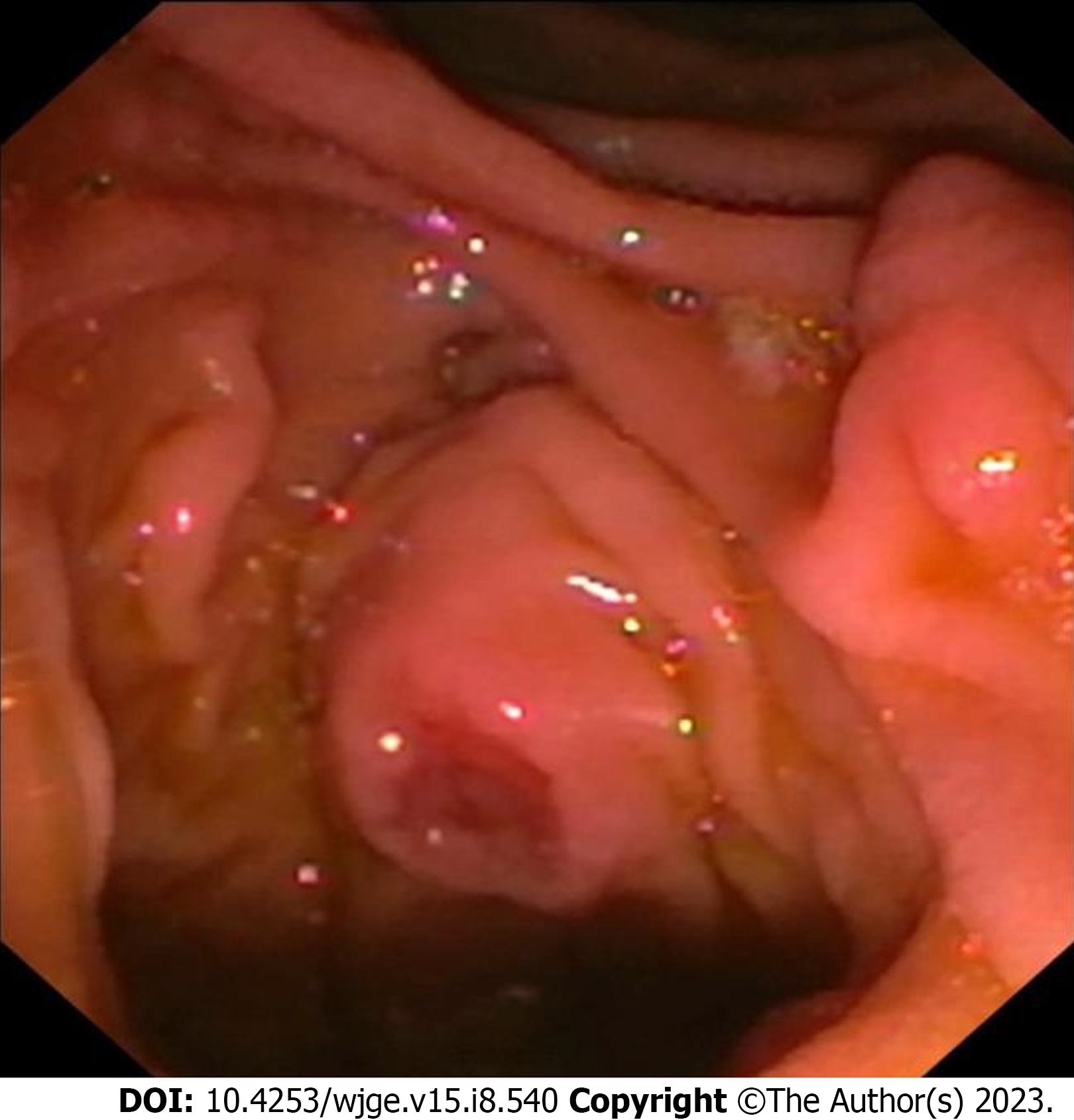
Figure 1
Image as visualized through a side-viewing dudenoscope showing an ulcerated papilla from which a biopsy was taken.
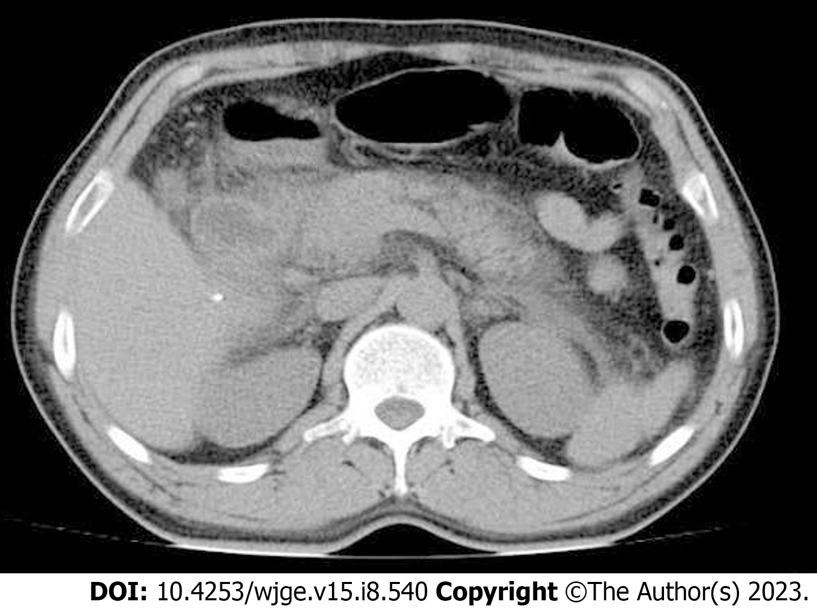
Figure 2
Computed tomography of the abdomen showing features consistent with acute pancreatitis such as pancreatic enlargement and diffuse peri-pancreatic fat stranding.
- Citation: George NM, Rajesh NA, Chitrambalam TG. Acute pancreatitis following endoscopic ampullary biopsy: A case report. World J Gastrointest Endosc 2023; 15(8): 540-544
- URL: https://www.wjgnet.com/1948-5190/full/v15/i8/540.htm
- DOI: https://dx.doi.org/10.4253/wjge.v15.i8.540


