Copyright
©The Author(s) 2023.
World J Gastrointest Endosc. Jun 16, 2023; 15(6): 420-433
Published online Jun 16, 2023. doi: 10.4253/wjge.v15.i6.420
Published online Jun 16, 2023. doi: 10.4253/wjge.v15.i6.420
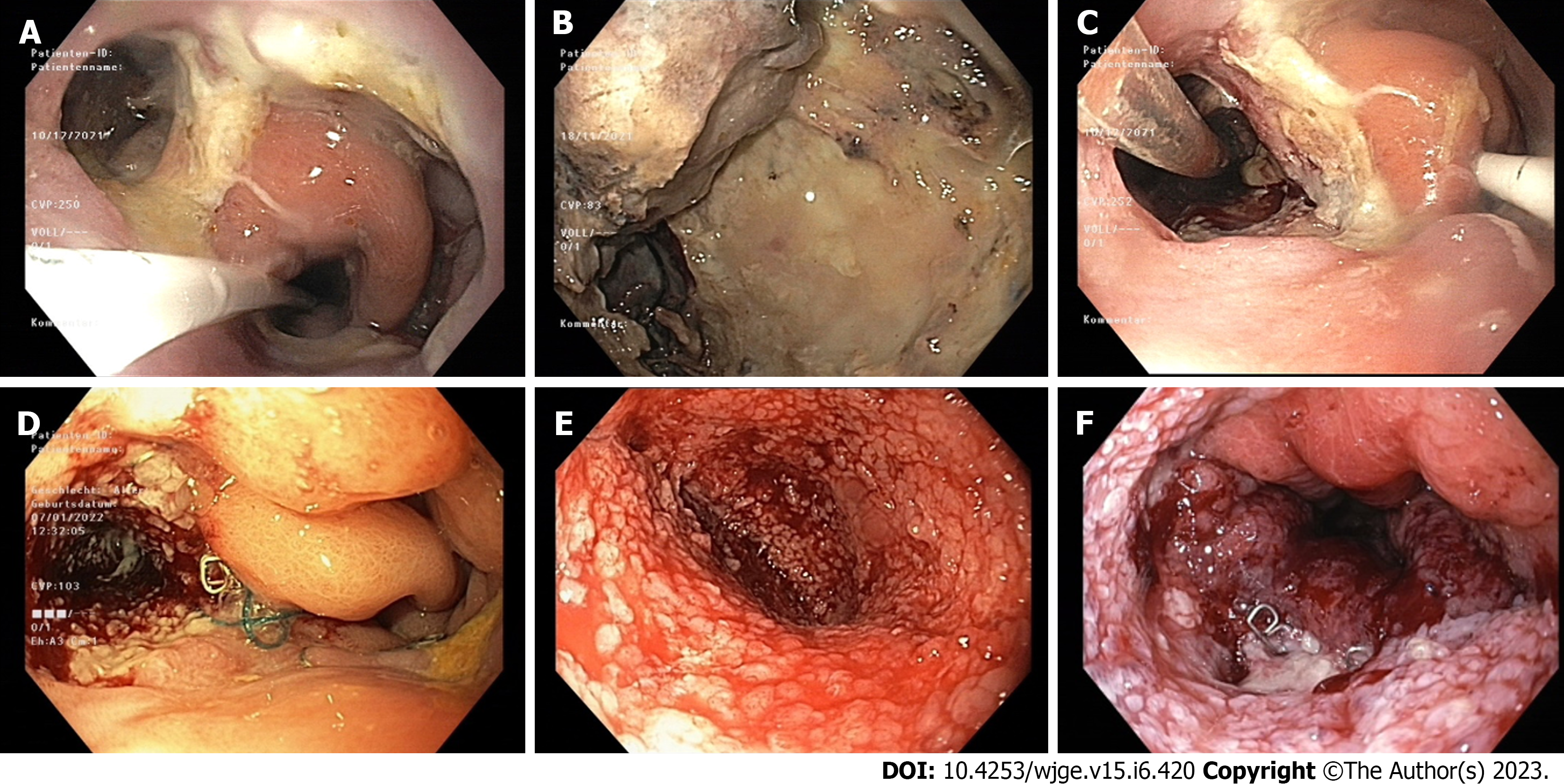
Figure 1 Intracavitary endoscopic vacuum therapy for an anastomotic leak after Ivor-Lewis esophagectomy.
A: 10 mm wide Anastomotic leak; B: 6 cm deep cavity with necrotic tissue, debris, and fibrin; C: Intracavitary positioning of an EsoSponge (a nasojejunal feeding tube is visible inside the lumen to the right); D: Downsizing of the defect after the first endoscopic vacuum therapy (EVT) session; E: Clean cavity with healthy granular tissue after the first EVT session; F: Healed anastomotic leak after 4 EVT sessions.
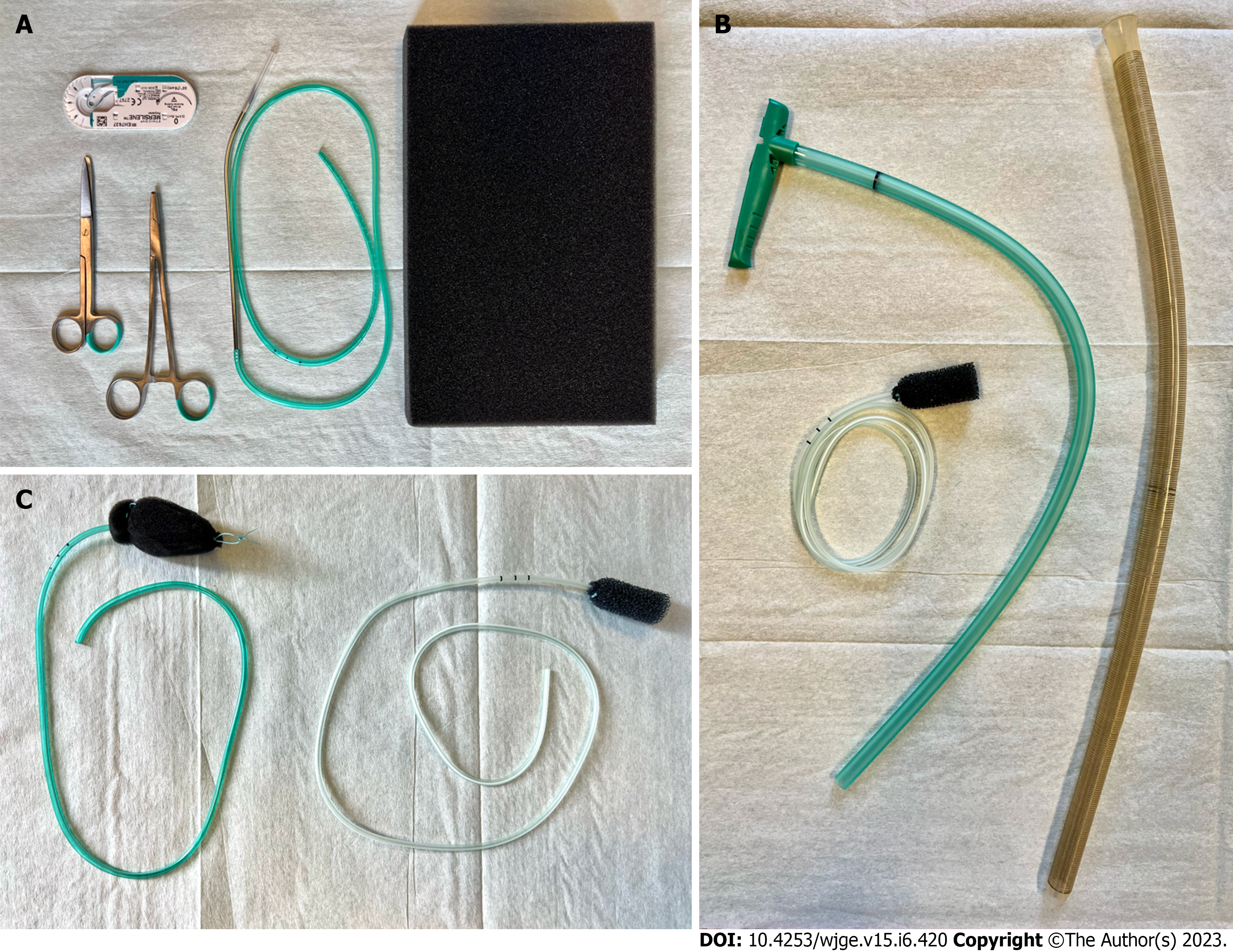
Figure 2 Endoscopic vacuum therapy materials.
A: The necessary materials for a handmade endoscopic vacuum therapy (EVT) sponge: Suture, scissors, needleholder, drain and polyurethane sponge; B: The components of the EsoSponge System: EVT sponge, pusher and overtube; C: A comparison of a handmade EVT sponge (left) and an EsoSponge (right).
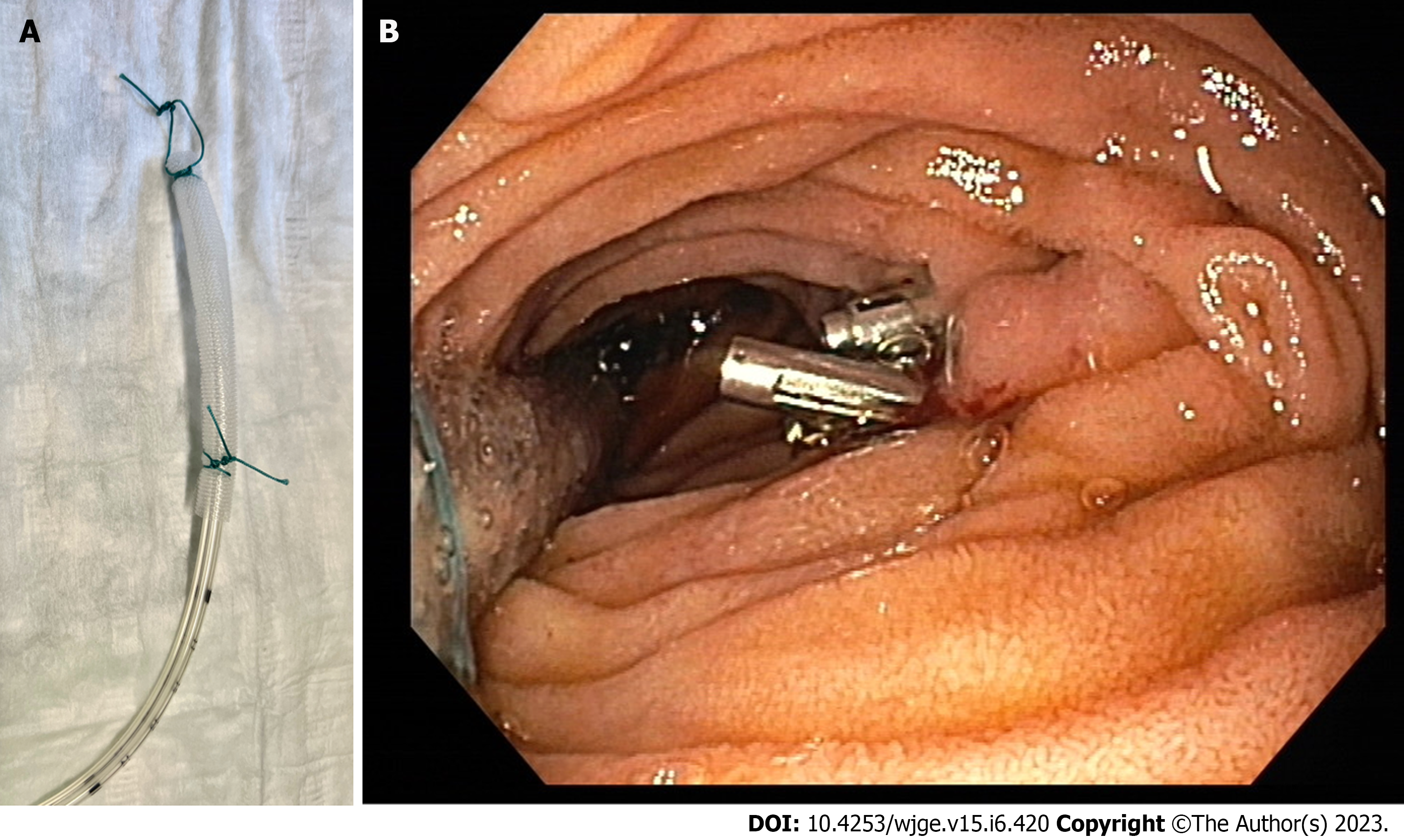
Figure 3 Open-pore film drainage.
A: The open-pore film drainage (OFD) system; B: Intraluminal application of OFD in the duodenum after a resection related perforation (clips are visible at the resection site).
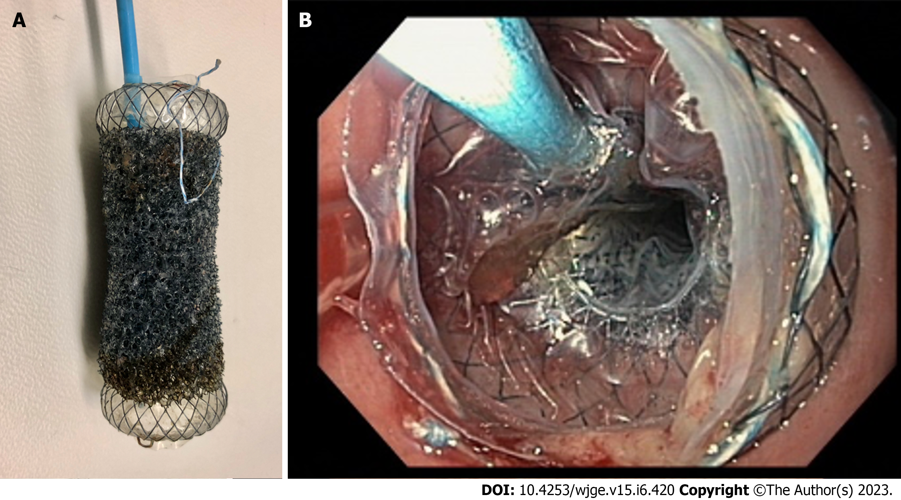
Figure 4 VAC-Stent (MICRO-TECH Europe GmbH, Düsseldorf, Germany).
A: The VAC-Stent system; B: The VAC-stent in place in the esophagus.
- Citation: Kouladouros K. Applications of endoscopic vacuum therapy in the upper gastrointestinal tract. World J Gastrointest Endosc 2023; 15(6): 420-433
- URL: https://www.wjgnet.com/1948-5190/full/v15/i6/420.htm
- DOI: https://dx.doi.org/10.4253/wjge.v15.i6.420


