Copyright
©The Author(s) 2020.
World J Gastrointest Endosc. Jul 16, 2020; 12(7): 198-211
Published online Jul 16, 2020. doi: 10.4253/wjge.v12.i7.198
Published online Jul 16, 2020. doi: 10.4253/wjge.v12.i7.198
Figure 1 Flow diagram for lesions examined and treatment.
EMR: Endoscopic mucosal resection; SM fibrosis: Sub-mucosal fibrosis.
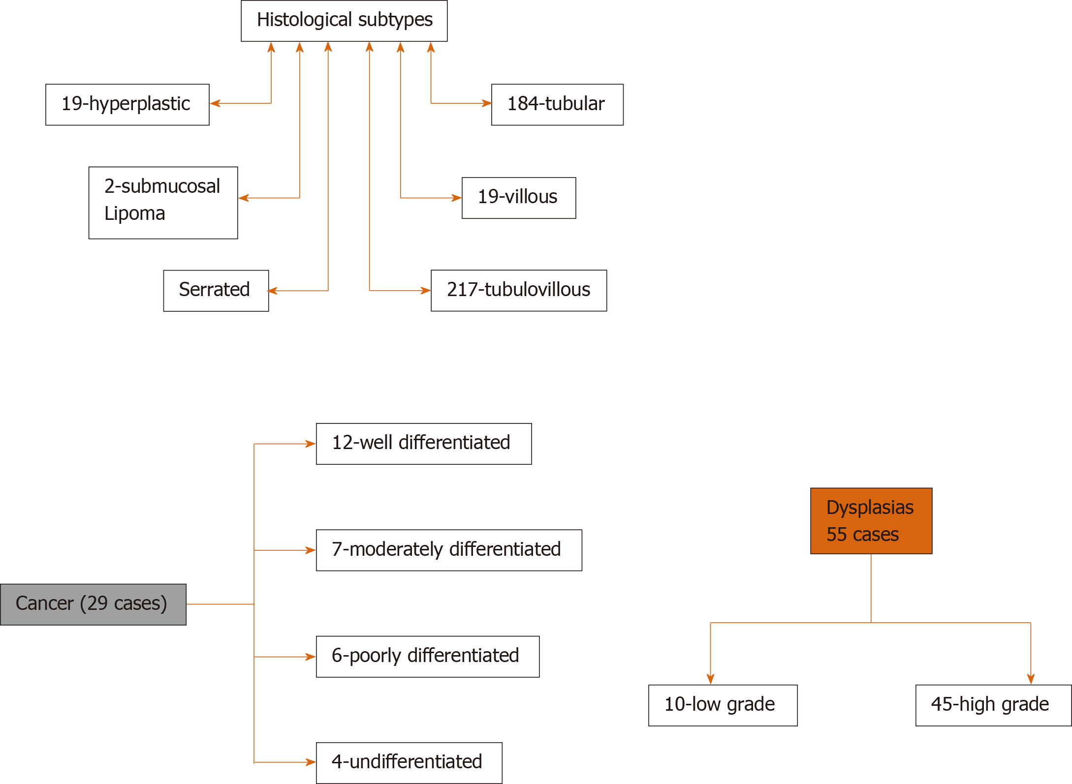
Figure 2 Histological subtypes and pathology results for the lesions.
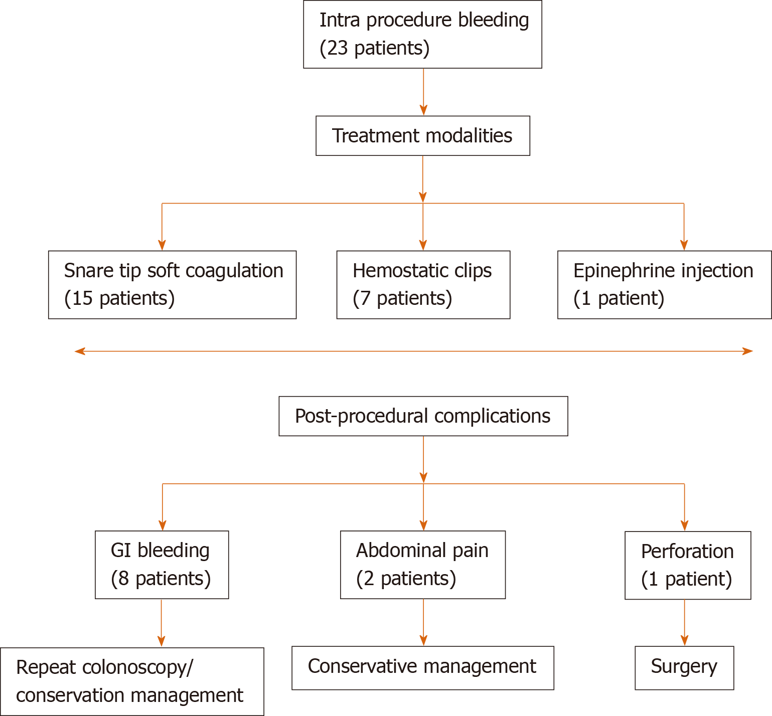
Figure 3 Complications and management of endoscopic mucosal resection.
Figure 4 large rectal polyp with steps of polyp injection and post resection area of the polyp.
A: Large rectal polyp; B: Steps of polyp injection; C: Post resection area of the polyp.
- Citation: Rashid MU, Khetpal N, Zafar H, Ali S, Idrisov E, Du Y, Stein A, Jain D, Hasan MK. Colon mucosal neoplasia referred for endoscopic mucosal resection: Recurrence of adenomas and prediction of submucosal invasion. World J Gastrointest Endosc 2020; 12(7): 198-211
- URL: https://www.wjgnet.com/1948-5190/full/v12/i7/198.htm
- DOI: https://dx.doi.org/10.4253/wjge.v12.i7.198


