Copyright
©The Author(s) 2020.
World J Gastrointest Endosc. Mar 16, 2020; 12(3): 83-97
Published online Mar 16, 2020. doi: 10.4253/wjge.v12.i3.83
Published online Mar 16, 2020. doi: 10.4253/wjge.v12.i3.83
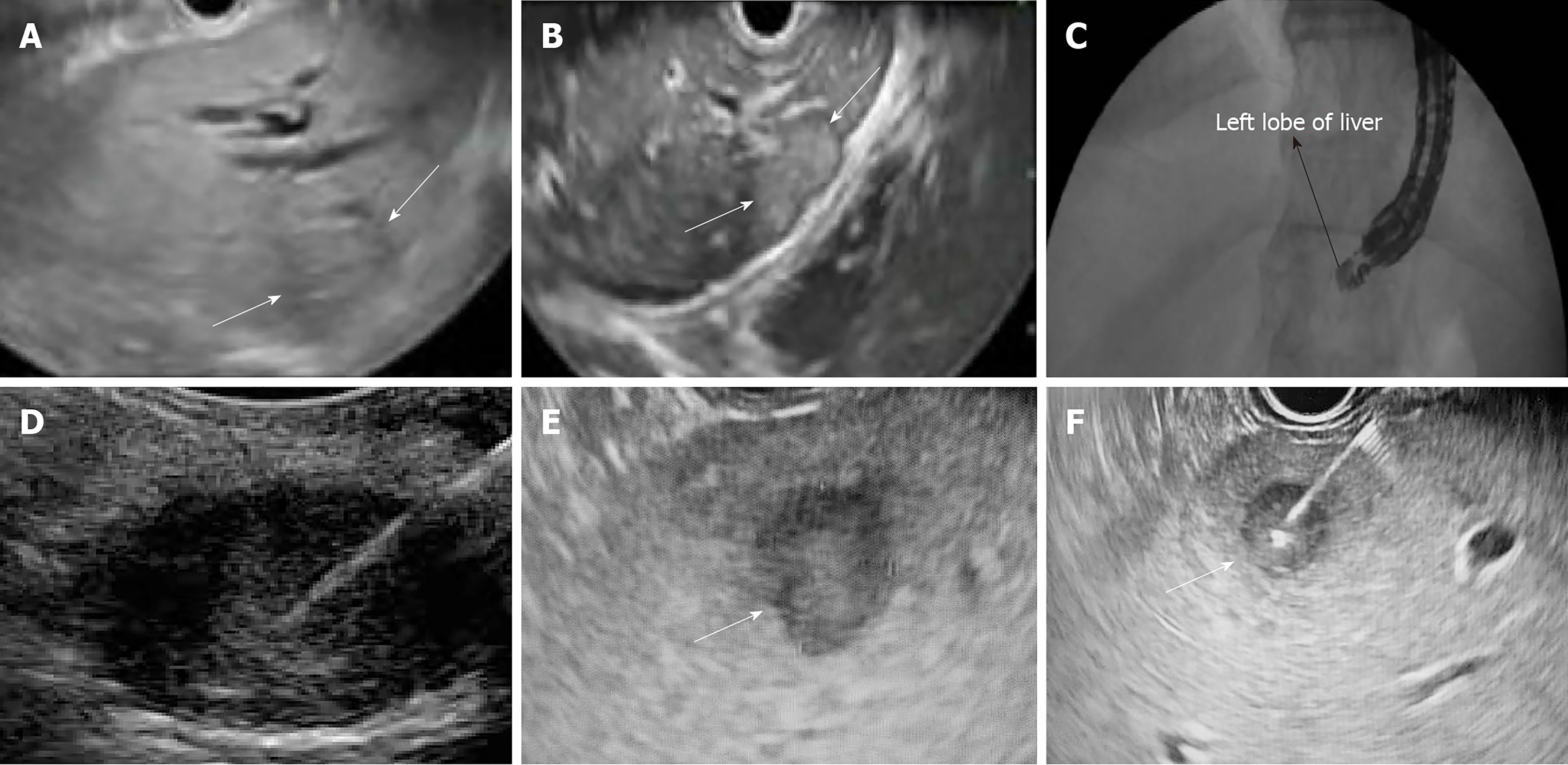
Figure 1 The endoscopic ultrasound guided liver biopsy provides clinicians with a real-time, detailed view of the biopsy needle through the course of the liver.
Figure 2 Histology of liver biopsy from the endoscopic ultrasound approach.
A: Liver parenchyma with macrovesicular steatosis and focal ballooning degeneration (200× magnification, hematoxylin-eosin staining); B: Liver parenchyma with portal tract (center, 200× magnification, hematoxylin-eosin staining); C: Liver biopsy performed to target a mass lesion that was a clinically suspected metastasis (40× magnification, hematoxylin-eosin staining). The majority of the biopsy is composed of pleomorphic epithelioid cells in sheets and trabeculae that was suggestive of metastatic germ cell tumor.
- Citation: Johnson KD, Laoveeravat P, Yee EU, Perisetti A, Thandassery RB, Tharian B. Endoscopic ultrasound guided liver biopsy: Recent evidence. World J Gastrointest Endosc 2020; 12(3): 83-97
- URL: https://www.wjgnet.com/1948-5190/full/v12/i3/83.htm
- DOI: https://dx.doi.org/10.4253/wjge.v12.i3.83


