Copyright
©The Author(s) 2019.
World J Gastrointest Endosc. Jul 16, 2019; 11(7): 438-442
Published online Jul 16, 2019. doi: 10.4253/wjge.v11.i7.438
Published online Jul 16, 2019. doi: 10.4253/wjge.v11.i7.438
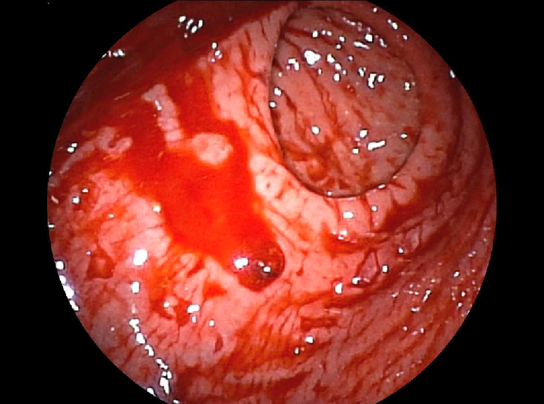
Figure 1 Endoscopic appearance of Dieulfoy's lesion.
A small pigmented protuberance with minimal surrounding erosion and no ulcerative lesion.
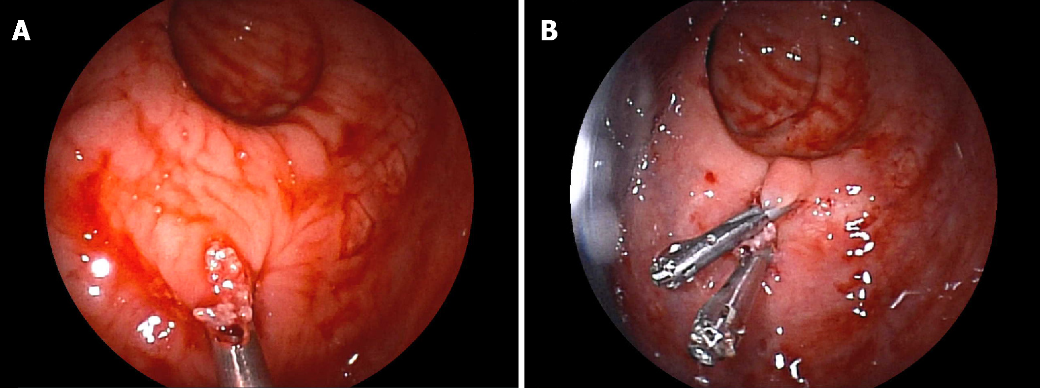
Figure 2 Endoscopic management of Dieulafoy’s lesion.
A: Placement of the first hemoclip; B: Placement of a second hemoclip to ensure hemostasis.
- Citation: Pineda-De Paz MR, Rosario-Morel MM, Lopez-Fuentes JG, Waller-Gonzalez LA, Soto-Solis R. Endoscopic management of massive rectal bleeding from a Dieulafoy's lesion: Case report. World J Gastrointest Endosc 2019; 11(7): 438-442
- URL: https://www.wjgnet.com/1948-5190/full/v11/i7/438.htm
- DOI: https://dx.doi.org/10.4253/wjge.v11.i7.438


