Copyright
©The Author(s) 2019.
World J Gastrointest Endosc. May 16, 2019; 11(5): 389-394
Published online May 16, 2019. doi: 10.4253/wjge.v11.i5.389
Published online May 16, 2019. doi: 10.4253/wjge.v11.i5.389
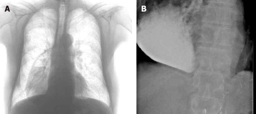
Figure 1 Chest x ray with a dilated gastric conduit with air fluid level and a contrast X ray showing meagre passage of contrast with dilated gastric conduit.
A: Chest X ray demonstration a dilated gastric conduit with air fluid level; B: Contrast X ray showing meagre passage of contrast with dilated gastric conduit.
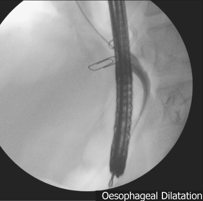
Figure 2 Bougie dilatation with two clip markers at the site of pyloric stricture.
Figure 3 Contract X ray deployed biodegradable stent with endoscope in the gastric conduit together injection catheter, endoscopic and outside view of the biodegradable stent.
A: Contrast in the deployed biodegradable (BD) stent with endoscope in the gastric conduit together injection catheter; B: Endoscopic view of the deployed BD stent; C: Sx Ella BD stent.
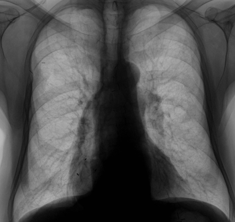
Figure 4 X ray showing well decompressed gastric conduit with proximal radio opaque markers of the biodegradable stent.
- Citation: Musbahi A, Viswanath Y. Post-oesophagectomy gastric conduit outlet obstruction following caustic ingestion, endoscopic management using a SX-ELLA biodegradable stent: A case report. World J Gastrointest Endosc 2019; 11(5): 389-394
- URL: https://www.wjgnet.com/1948-5190/full/v11/i5/389.htm
- DOI: https://dx.doi.org/10.4253/wjge.v11.i5.389


