Copyright
©The Author(s) 2019.
World J Gastrointest Endosc. Mar 16, 2019; 11(3): 249-255
Published online Mar 16, 2019. doi: 10.4253/wjge.v11.i3.249
Published online Mar 16, 2019. doi: 10.4253/wjge.v11.i3.249
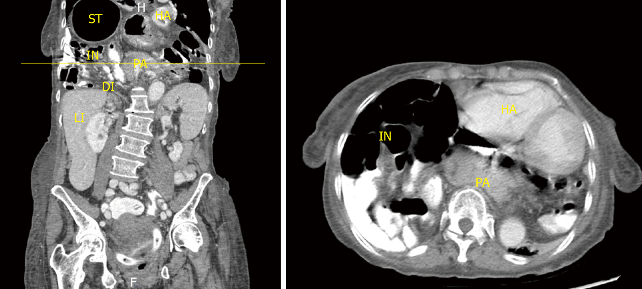
Figure 1 Demonstrate the contrast-enhanced computed tomography (oral and intravenous), coronal and axial reconstruction with large hiatal hernia with pancreas in the chest.
ST: Stomach; IN: Small intestine; LI: Large intestine; PA: Pancreas; DI: Diaphragm; HA: Heart.
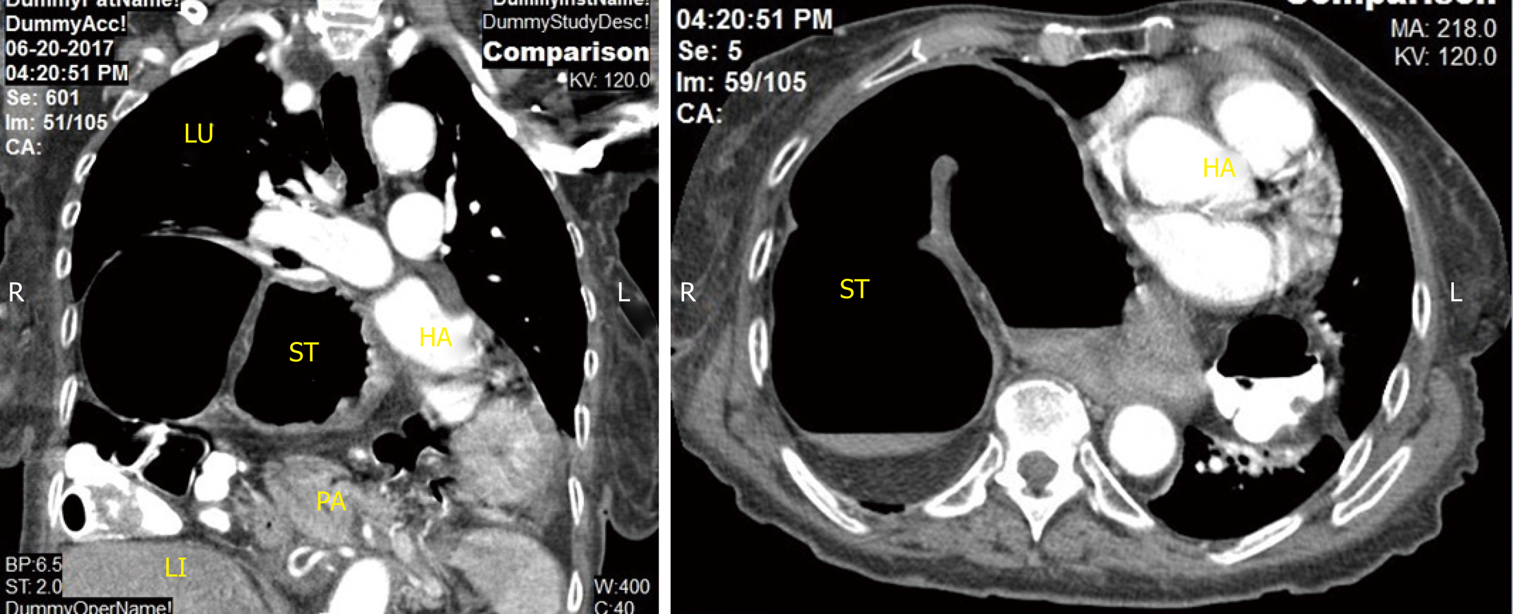
Figure 2 Demonstrate the computed tomography of the chest with large hiatal hernia.
No pancreas in the hernia sac. ST: Stomach; LI: Large intestine; PA: Pancreas; HA: Heart; LU: Lung.
- Citation: Kamal MU, Baiomi A, Erfani M, Patel H. Rare sequalae of hiatal hernia causing pancreatitis and hepatitis: A case report. World J Gastrointest Endosc 2019; 11(3): 249-255
- URL: https://www.wjgnet.com/1948-5190/full/v11/i3/249.htm
- DOI: https://dx.doi.org/10.4253/wjge.v11.i3.249


