Copyright
©The Author(s) 2018.
World J Gastrointest Endosc. Jul 16, 2018; 10(7): 125-129
Published online Jul 16, 2018. doi: 10.4253/wjge.v10.i7.125
Published online Jul 16, 2018. doi: 10.4253/wjge.v10.i7.125
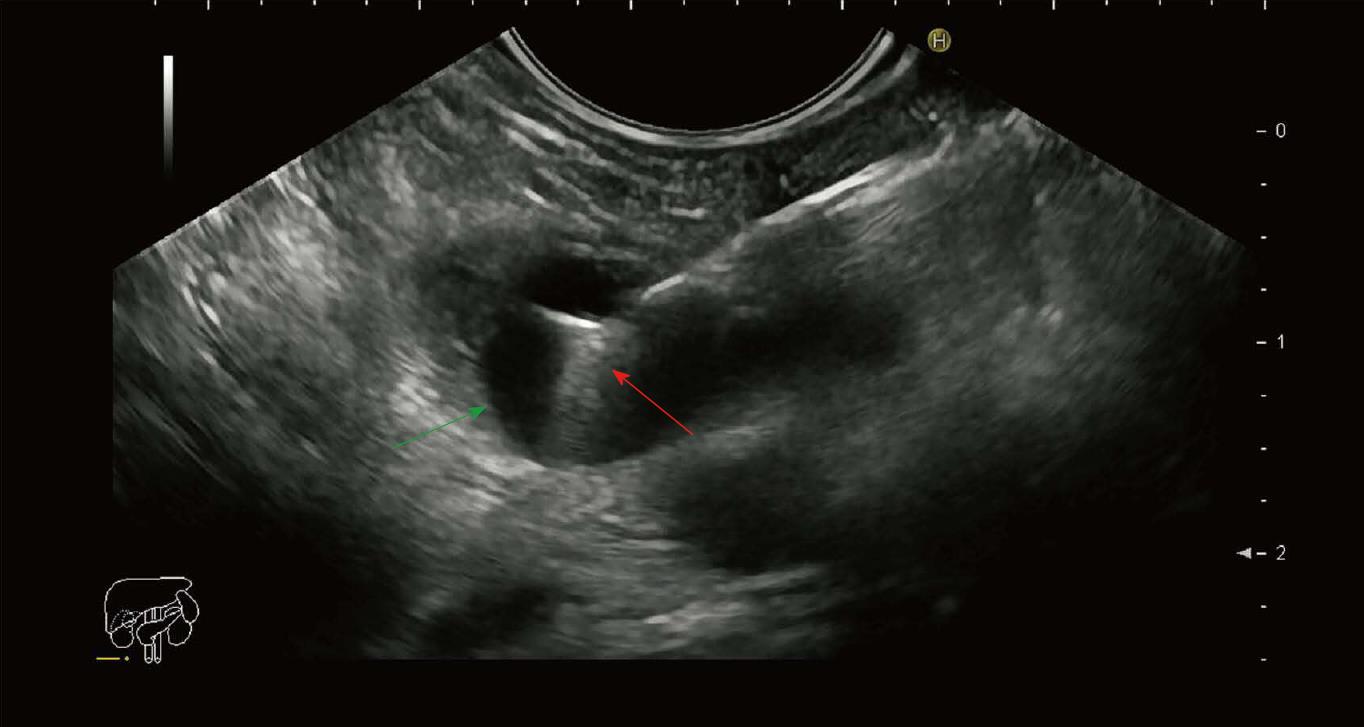
Figure 1 Endoscopic ultrasound image of the pancreatic cyst punctured with a 19 gauge needle with a microbiopsy forceps.
Green arrow: Cyst wall; Red arrow: Microbiopsy forceps.
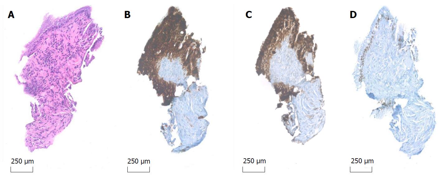
Figure 2 Microbiopsy specimen 20 × original magnification.
A: Hematoxylin and eosin stain (A) reveals fragments of mucinous epithelium with goblet cells and basally oriented nuclei; B-D: The epithelial cells are immunohistochemical positive for MUC1 (B), MUC5AC (C) and focal positive for CDX2 (D), indicative of IPMN of mixed type: Pancreatobiliary and intestinal.
- Citation: Rift CV, Kovacevic B, Karstensen JG, Plougmann J, Klausen P, Toxværd A, Kalaitzakis E, Hansen CP, Hasselby JP, Vilmann P. Diagnosis of intraductal papillary mucinous neoplasm using endoscopic ultrasound guided microbiopsies: A case report. World J Gastrointest Endosc 2018; 10(7): 125-129
- URL: https://www.wjgnet.com/1948-5190/full/v10/i7/125.htm
- DOI: https://dx.doi.org/10.4253/wjge.v10.i7.125


