Copyright
©2014 Baishideng Publishing Group Inc.
World J Hepatol. Aug 27, 2014; 6(8): 580-595
Published online Aug 27, 2014. doi: 10.4254/wjh.v6.i8.580
Published online Aug 27, 2014. doi: 10.4254/wjh.v6.i8.580
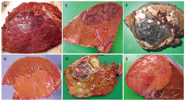
Figure 5 Hepatocellular adenoma macroscopic aspects of different subtypes: A-C: Examples of HNF1A mutated hepatocellular adenoma (H-HCA).
A: Woman born in 1972; oral contraceptives for 8 years. Discovery of a liver nodule (3.5 cm). No regression after stopping contraceptives. Fear of complications by the patient. Left hepatectomy in 2008; fresh specimen: yellowish tumor, clearer than the surrounding liver. The diagnosis of H-HCA is likely but not self-evident. The diagnosis was confirmed by immunohistochemistry. B: Woman born in 1974; oral contraceptives for 12 years. Liver nodules discovered by chance on imaging and diagnosis of adenomatosis. Largest nodule 5 cm. Segmentectomy IVb 2006. Other small nodules. One nodule was an focal nodular hyperplasia (FNH). Fixed specimen of one nodule: yellowish, clear tumor, non encapsulated, contrasting with the surrounding liver. The diagnosis of H-HCA is likely. The diagnosis was confirmed by immunohistochemistry. C: Woman born in 1978. Massive right hepatomegaly discovered in the obstetric department (miscarriage at 6 wk). No oral contraception. Right hepatectomy in 2004. Fresh specimen: large irregular, mammillated tumor occupying the whole right liver. This is an exceptional case. The diagnosis was confirmed by immunohistochemistry. D-F: Examples of IHCA (D, E) and β-IHCA (F). D: Woman born in 1968; overweight, BMI > 40 kg/m2. Oral contraceptives for 18 years. Several nodules detected on imaging. Doubtful diagnosis. Surgical biopsies: HCA. Right hepatectomy in 2008 (largest nodule 10 cm). In 2009, another known HCA removed in the left liver (11 cm). Fresh specimen: reddish tumor with congestive areas. The diagnosis of inflammatory HCA (IHCA) is likely. The diagnosis was confirmed by immunohistochemistry. E: Woman born in 1956; oral contraceptives for 31 years. Biological abnormalities (inflammatory syndrome). CT scan and MRI multiple liver nodules (largest nodule 7 cm) in favor of IHCA. Surgery in 2007: bisegmentectomy VI-VII plus 2 tumorectomies. Fresh specimen: ill defined tumor with congestive strands. The diagnosis of IHCA is very likely. The diagnosis was confirmed by immunohistochemistry. F: Woman born in 1971; liver hemorrhage. Imaging: 5 nodules, largest 8 cm. No oral contraceptives; BMI 20.4 kg/m2. Segmentectomy III, VI, VIII 2007. Fixed specimen: large hematoma and a narrow viable tissue at the periphery. No obvious diagnosis. The diagnosis of HCC cannot be ruled out. The diagnosis was confirmed by immunohistochemistry. G: Example of β-catenin HCA. Woman born in 1981; one nodule 8 cm discovered by chance. Imaging HCA. Oral contraceptives for 8 years; BMI 21.1 kg/m2. Right hepatectomy 2005. Fresh specimen: well limited clear nodule. The diagnosis of HCA is likely. H-HCA and IHCA are unlikely. H: Example of HCC developed on β-IHCA. Woman born in 1934; intramuscular injection of hormones as contraceptive. Liver nodule interpreted as hemangioma, known for several years. Growth of the nodule. Segmentectomy in 2000. Fresh specimen: irregular, multinodular tumor, with a large necrotic and hemorrhagic area ( arrowhead) surrounded by a fibrous rim. The diagnosis of HCC is likely. I: Example of unclassified HCA. Woman born in 1983; abdominal pain. Imaging: one nodule 8 cm; no final diagnosis. Oral contraceptives for 8 years. BMI 20.2 kg/m2. Right hepatectomy 2007. Fresh specimen: well limited clear nodule with a pale reddish area. The diagnosis of HCA is likely. H-HCA and IHCA are unlikely.
- Citation: Sempoux C, Balabaud C, Bioulac-Sage P. Pictures of focal nodular hyperplasia and hepatocellular adenomas. World J Hepatol 2014; 6(8): 580-595
- URL: https://www.wjgnet.com/1948-5182/full/v6/i8/580.htm
- DOI: https://dx.doi.org/10.4254/wjh.v6.i8.580


