Copyright
©The Author(s) 2017.
World J Hepatol. Feb 8, 2017; 9(4): 171-179
Published online Feb 8, 2017. doi: 10.4254/wjh.v9.i4.171
Published online Feb 8, 2017. doi: 10.4254/wjh.v9.i4.171
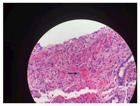
Figure 1 Spindle cells with cytokeratin 7 staining positive.
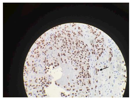
Figure 2 Positive human herpersvirus-8 immunohistochemical staining.
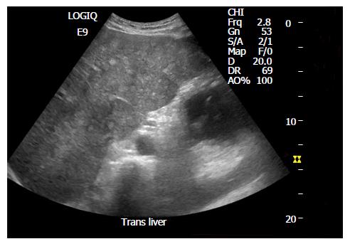
Figure 3 Ultrasound image with multiple small round hyperechoic nodules.
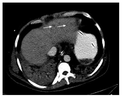
Figure 4 Computerized tomography scan enlarged inhomogeneous liver with multiple hypodense lesions.
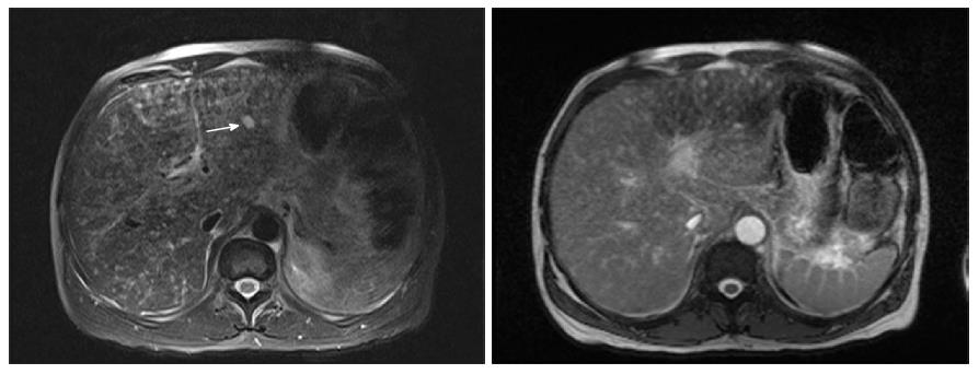
Figure 5 SPAIR and T2 images Kaposi sarcoma on magnetic resonance imaging.
- Citation: Van Leer-Greenberg B, Kole A, Chawla S. Hepatic Kaposi sarcoma: A case report and review of the literature. World J Hepatol 2017; 9(4): 171-179
- URL: https://www.wjgnet.com/1948-5182/full/v9/i4/171.htm
- DOI: https://dx.doi.org/10.4254/wjh.v9.i4.171


