Copyright
©The Author(s) 2017.
World J Hepatol. Aug 8, 2017; 9(22): 973-978
Published online Aug 8, 2017. doi: 10.4254/wjh.v9.i22.973
Published online Aug 8, 2017. doi: 10.4254/wjh.v9.i22.973
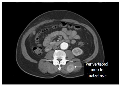
Figure 1 Computed tomography of the recurrent tumor.
Computed tomography scan showing an enhancing lesion 3.7 cm in size in the left paravertebral musculature.
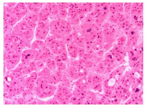
Figure 2 Histology of the paravertebral muscle tumor.
A biopsy of the mass showed moderate-to-poorly differentiated hepatocellular carcinoma (HCC), consistent with metastatic HCC (hematoxylin and eosin, × 200).
- Citation: Takahashi K, Putchakayala KG, Safwan M, Kim DY. Extrahepatic metastasis of hepatocellular carcinoma to the paravertebral muscle: A case report. World J Hepatol 2017; 9(22): 973-978
- URL: https://www.wjgnet.com/1948-5182/full/v9/i22/973.htm
- DOI: https://dx.doi.org/10.4254/wjh.v9.i22.973


