Copyright
©The Author(s) 2017.
World J Hepatol. Jul 28, 2017; 9(21): 930-944
Published online Jul 28, 2017. doi: 10.4254/wjh.v9.i21.930
Published online Jul 28, 2017. doi: 10.4254/wjh.v9.i21.930
Figure 1 Graft with V5 to be anastomosed with recipient liver transplantation hepatic vein.
A: Computed tomography venography showing large V5; B: A jumping graft between the V5 vein of liver graft and liver transplantation hepatic vein of recipient.
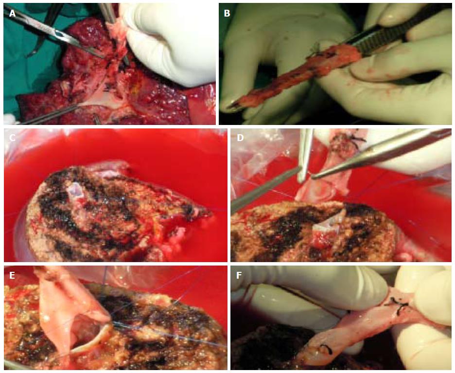
Figure 2 Graft with V8 to be anastomosed with recipient inferior vena cava by jumping graft.
A: Obtaining the venous graft from native PV; B: The venous graft; C: V8 vein during back table preparation; D and E: Anastomosing the venous graft to V8; F: Preparation for anastomosing the venous graft to IVC. IVC: Inferior vena cava.
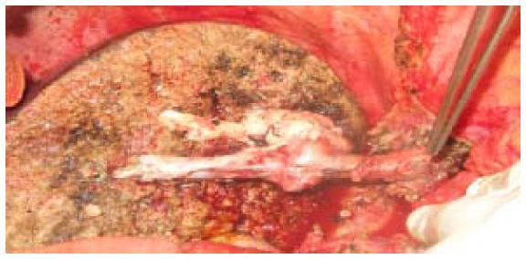
Figure 3 Venous graft obtained from native PUV, portal venous and hepatic vein to communicate 2 V5, 1V8 and right hepatic vein of liver graft with recipient inferior vena cava.
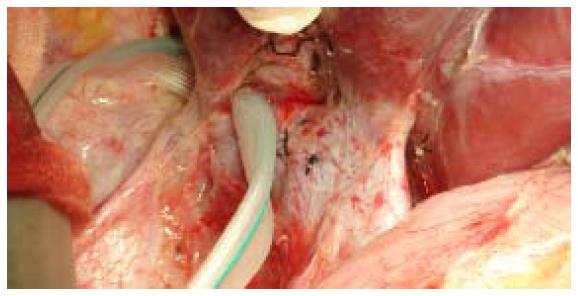
Figure 4 Small right hepatic vein (encircled) and large right inferior vein harvested and anastomosed with recipient inferior vena cava.
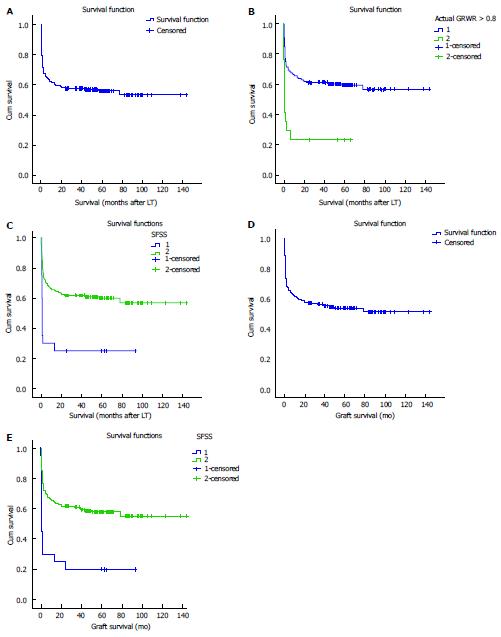
Figure 5 Kaplan-Meier survival curves (1, 2, 3).
A: KM survival curve; B: SFSG and survival [SFSG (GRWR < 0.8) = 2, Log-Rank = 0.00]; C: SFSS and survival (SFSS = 1, Log-Rank = 0.00); D: KM graft survival curve; E: SFSS and graft survival (SFSS = 1, Log-Rank = 0.00). SFSG: Small for size graft; GRWR: Graft recipient weight ratio; SFSS: Small for size syndrome.
- Citation: Shoreem H, Gad EH, Soliman H, Hegazy O, Saleh S, Zakaria H, Ayoub E, Kamel Y, Abouelella K, Ibrahim T, Marawan I. Small for size syndrome difficult dilemma: Lessons from 10 years single centre experience in living donor liver transplantation. World J Hepatol 2017; 9(21): 930-944
- URL: https://www.wjgnet.com/1948-5182/full/v9/i21/930.htm
- DOI: https://dx.doi.org/10.4254/wjh.v9.i21.930


