Copyright
©The Author(s) 2016.
World J Hepatol. Dec 18, 2016; 8(35): 1576-1583
Published online Dec 18, 2016. doi: 10.4254/wjh.v8.i35.1576
Published online Dec 18, 2016. doi: 10.4254/wjh.v8.i35.1576
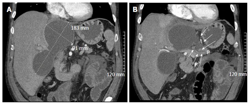
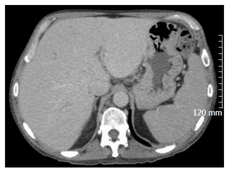
Figure 1 Abdominal computed tomography images showing bilobed intrahepatic pancreatic pseudocysts (A), including connection to main pancreatic duct (B, arrows).
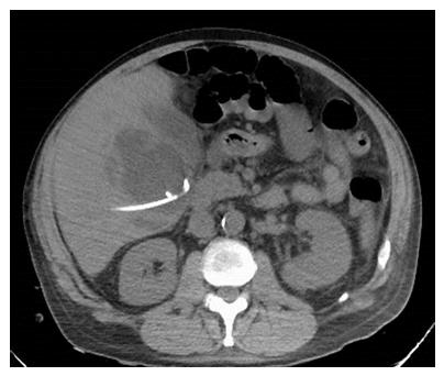
Figure 2 Abdominal computed tomography image showing percutaneous transhepatic drainage of the more superficial, inferior lobe.
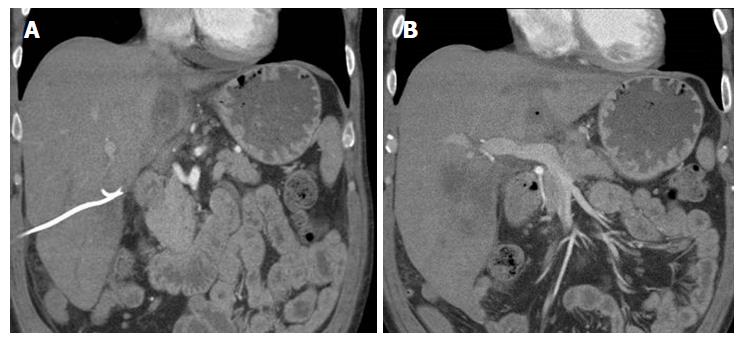
Figure 3 Abdominal computed tomography images showing the superficial, inferior lobe to be well drained with the pigtail in place (A), but the deeper superior collection containing a small bubble of gas (B).
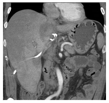
Figure 4 Abdominal computed tomography image showing drain was repositioned into the deeper lobe seen in Figure 3B.
Figure 5 Abdominal computed tomography image showing resolution of the intrahepatic pancreatic pseudocysts at 3 mo following the initial intervention.
- Citation: Demeusy A, Hosseini M, Sill AM, Cunningham SC. Intrahepatic pancreatic pseudocyst: A review of the world literature. World J Hepatol 2016; 8(35): 1576-1583
- URL: https://www.wjgnet.com/1948-5182/full/v8/i35/1576.htm
- DOI: https://dx.doi.org/10.4254/wjh.v8.i35.1576


