Copyright
©The Author(s) 2016.
World J Hepatol. Nov 28, 2016; 8(33): 1442-1451
Published online Nov 28, 2016. doi: 10.4254/wjh.v8.i33.1442
Published online Nov 28, 2016. doi: 10.4254/wjh.v8.i33.1442
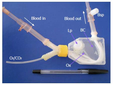
Figure 1 Minibioreactor device.
BC: Biological compartment; Lp: Loading port; O2/CO2: Carbogen supply line; Ox: Silicone tube oxygenator; Bsp: Blood sample port.
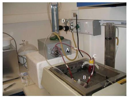
Figure 2 In vitro perfusion system.
A: Peristaltic pump; B: Minibioreactor; C: Blood reservoir; D: External oxygen supply.
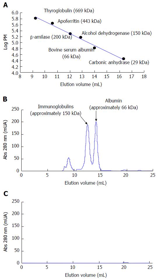
Figure 3 Protein analysis by gel filtration chromatography.
A: Calibration curve carried out with molecular weight markers; B: Chromatogram obtained for a sample of basal plasma; C: Chromatogram obtained for a sample of biological compartment, taken after 120 min of perfusion n = 5.
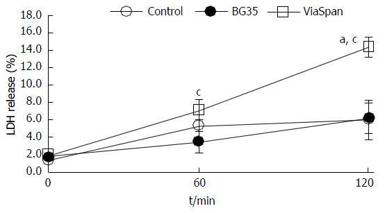
Figure 4 Time course of lactate dehydrogenase release during 120 min of minibioreactor perfusion determined for fresh and cold preserved liver microorgans in BG35 and ViaSpan® solutions.
Data are expressed as mean ± SD for 6 liver microorgans (LMOs) preparations. Different from control, aP < 0.05; Different from all the other reoxygenation times, cP < 0.05, n = 6 LMOs independent preparations for each condition. LDH: Lactate dehydrogenase.
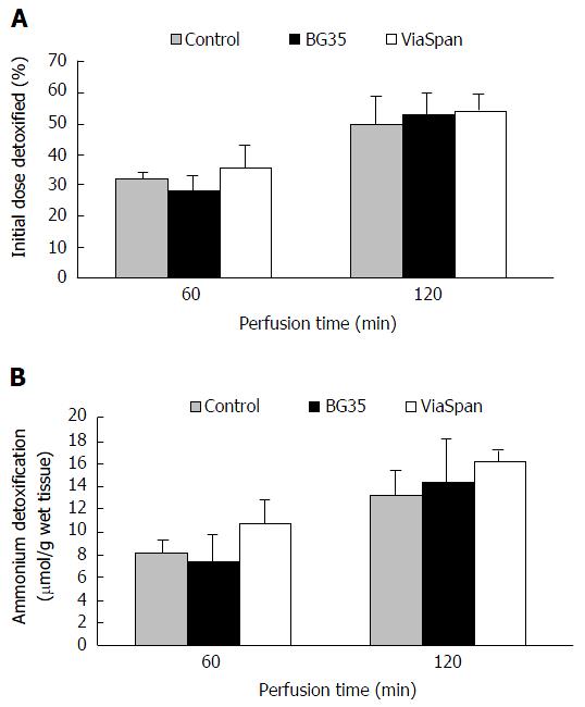
Figure 5 Evolution of (A) initial dose of ammonium detoxified (%) (B) detoxification of ammonia (μmol/g wet tissue) for fresh cold preserved liver Microorgans in BG35 and ViaSpan® solutions used as a biological component in the minibioreactor.
Data are expressed as mean ± SD, n = 6 liver Microorgans independent preparations for each condition.
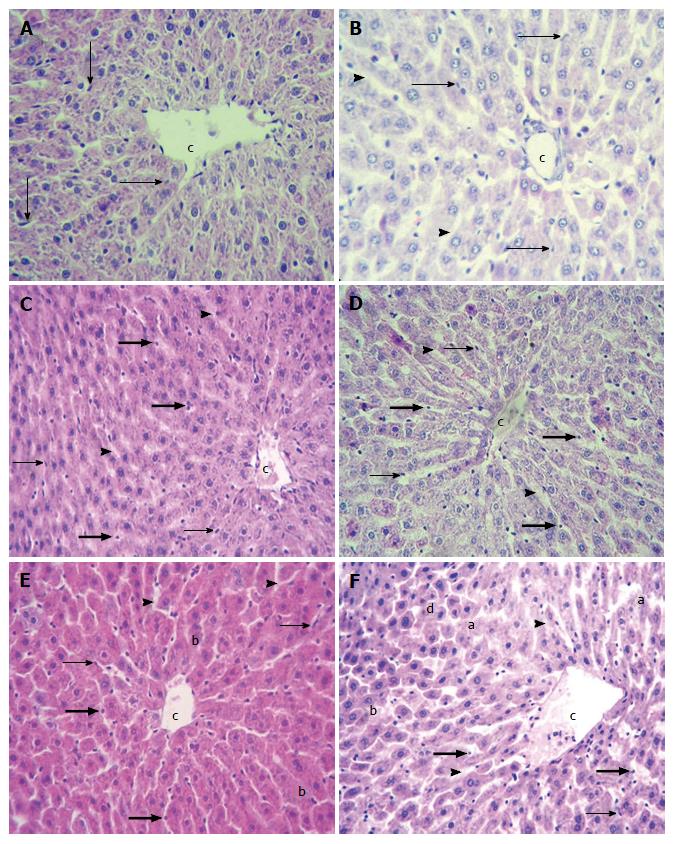
Figure 6 Liver microorgans histology.
Hematoxylin-eosin. Samples were taken from control and preserved groups at the beginning (A, C and E) and at the end (t = 120 min) (B, D and F) of the experiment. Controls (A and B) showed normal hepatic parenchyma with fusiform endothelial cells (arrows) attached to perisinusoidal extracellular matrix and conserved hepatocyte cords. Sinusoids appeared dilated (bold arrow head) after 120 min. Liver microorgans (LMOs) preserved in BG35 (C and D) presented conserved hepatic architecture with fusiform (arrows) and rounded (bold arrows) endothelial cells. Sinusoids were dilated (bold arrow heads). LMOs preserved in ViaSpan® (E and F) had fusiform (arrows) and rounded (bold arrows) endothelial cells, dilated sinusoids (bold arrow head), and balonized hepatocytes (b) at the beginning of the experiment. F: After 120 min, blebs (a) and areas of hepatocyte trabecular disruption (d) were also found. Central Vein (CV). Magnification × 200. n = 6 LMOs independent preparations for each condition. c: Central vein.
- Citation: Pizarro MD, Mediavilla MG, Quintana AB, Scandizzi ÁL, Rodriguez JV, Mamprin ME. Performance of cold-preserved rat liver Microorgans as the biological component of a simplified prototype model of bioartificial liver. World J Hepatol 2016; 8(33): 1442-1451
- URL: https://www.wjgnet.com/1948-5182/full/v8/i33/1442.htm
- DOI: https://dx.doi.org/10.4254/wjh.v8.i33.1442


