Copyright
©The Author(s) 2016.
World J Hepatol. Nov 18, 2016; 8(32): 1414-1418
Published online Nov 18, 2016. doi: 10.4254/wjh.v8.i32.1414
Published online Nov 18, 2016. doi: 10.4254/wjh.v8.i32.1414
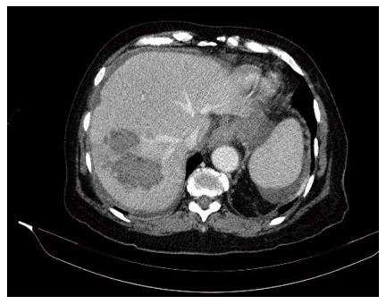
Figure 1 Computed tomography scan of the liver.
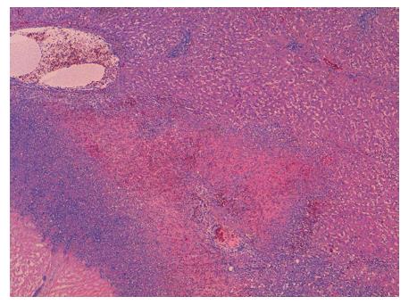
Figure 2 Liver (hematoxylin and eosin; × 40 original magnification): Bleeding, abscesses and avascular necrosis.
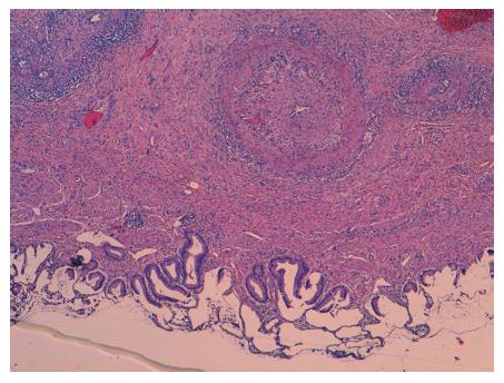
Figure 3 Gallbladder (hematoxylin and eosin; × 40 original magnification): Acute vasculitis.
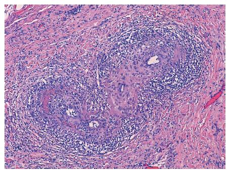
Figure 4 Gallbladder (hematoxylin and eosin; × 100 original magnification): Acute vasculitis with fibrinoid necrosis in muscular arteries of parietal medium caliber.
- Citation: Gómez-Luque I, Alconchel F, Ciria R, Ayllón MD, Luque A, Sánchez M, López-Cillero P, Briceño J. Spontaneous liver rupture as first sign of polyarteritis nodosa. World J Hepatol 2016; 8(32): 1414-1418
- URL: https://www.wjgnet.com/1948-5182/full/v8/i32/1414.htm
- DOI: https://dx.doi.org/10.4254/wjh.v8.i32.1414


