Copyright
©The Author(s) 2016.
World J Hepatol. Apr 8, 2016; 8(10): 461-470
Published online Apr 8, 2016. doi: 10.4254/wjh.v8.i10.461
Published online Apr 8, 2016. doi: 10.4254/wjh.v8.i10.461
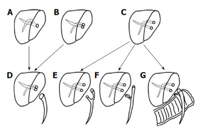
Figure 1 Types of biliary anastomoses and corresponding biliary reconstructions[54].
A: Single duct anastomosis; B: Double duct - minimum distance between two donor ducts, requires ductoplasty with recipient CBD; C: Double duct - two donor duct are far away, requires two separate duct anastomosis or a hepaticojejunostomy; D: Single duct to duct reonstruction; E: Double duct to duct reconstruction using right and left hepatic ducts; F: Double duct to duct reconstruction using cystic and CHD; G: Mixed type using duct to duct and hepaticojejunostomy. CHD: Common hepatic duct; CBD: Common bile duct.
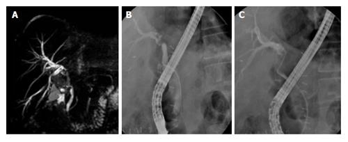
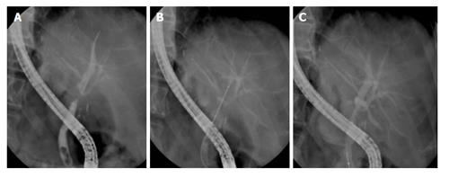
Figure 2 Anastomotic stricture - single duct anastomosis.
A: Magnetic resonance cholangiopancreatography shows stricture at the anastomotic site of a single duct anastomosis; B: Endoscopic retrograde cholangiopancreatography (ERCP) in the same patient shows the stricture; C: ERCP in same patient shows guide wire negotiated across the stricture.
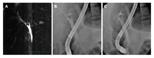
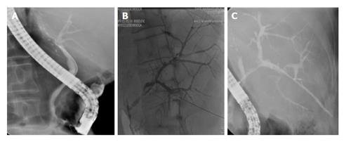
Figure 3 Anastomotic stricture - double duct anastomosis.
A: Magnetic resonance cholangiopancreatography image shows stricture across both RASD as well as RPSD ductal anastomosis; B: Endoscopic retrograde cholangiopancreatography (ERCP) image shows guide wire negotiated across RPSD in this patient; C: ERCP image shows guidewire negotiated across RASD in this patient.
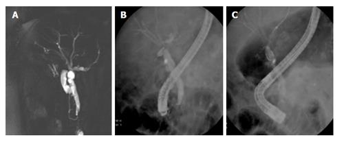
Figure 4 Anastomotic stricture - ductoplasty.
A: Magnetic resonance cholangiopancreatography image of a ductoplasty of RASD and RPSD to common hepatic duct; B: Endoscopic retrograde cholangiopancreatography (ERCP) image shows stricture at ductoplasty site; C: ERCP image shows guide wire across one ductal system.
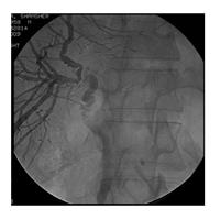
Figure 5 Anastomotic stricture - cystic duct anastomosis (endoscopic retrograde cholangiopancreatography failed, patient underwent percutaneous transhepatic biliary drainage).
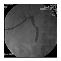
Figure 6 Cystic duct anastomosis after dilatation.
This patient developed stricture again and underwent a hepaticojejunostomy.
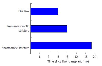
Figure 7 Timeline of biliary complications after transplant.
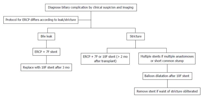
Figure 8 Protocol for endoscopic intervention (please see text also).
ERCP: Endoscopic retrograde cholangiopancreatography.
Figure 9 Balloon dilatation of biliary stricture.
A: Endoscopic retrograde cholangiopancreatography (ERCP) images show stricture at the anastomotic site; B: ERCP image showing balloon dilatation of the stricture; C: Successful obliteration of the waist of stricture after balloon dilatation.
Figure 10 Rendezvous procedure.
A: Endoscopic retrograde cholangiopancreatography opacified only RASD; B: RPSD accessed via percutaneous transhepatic biliary drainage; C: Rendezvous procedure being performed.
- Citation: Wadhawan M, Kumar A. Management issues in post living donor liver transplant biliary strictures. World J Hepatol 2016; 8(10): 461-470
- URL: https://www.wjgnet.com/1948-5182/full/v8/i10/461.htm
- DOI: https://dx.doi.org/10.4254/wjh.v8.i10.461


