Copyright
©The Author(s) 2016.
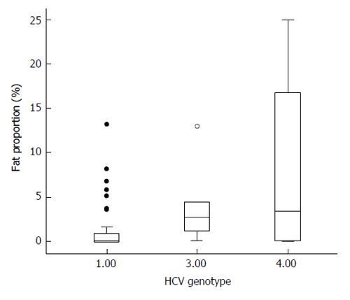
Figure 1 Fat amount among the three hepatitis C virus genotypes included in this study.
Patients with genotype 1 (the most frequent) show significantly less amount of fat than patients affected by genotype 3 or 4 (solid circles are outliers, and hollow circle, extreme values). HCV: Hepatitis C virus.
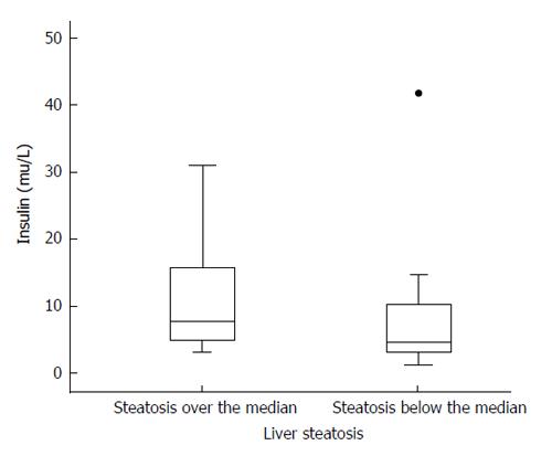
Figure 2 Serum insulin levels among patients with liver steatosis over the median (left) and below the median (right).
Differences are not statistically significant, but there is a trend to higher values among patients with intense steatosis (P = 0.059). Solid circle represents an outlier.
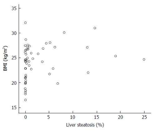
Figure 3 Scattergram showing the relation of body mass index with the amount of liver steatosis.
Despite a significant relationship between both variables (ρ = 0.41; P = 0.002), as shown, some patients with BMI over 30 show no steatosis at all or only minimal amount of liver fat, in contrast with some others with BMI below 25 and marked steatosis in their livers. BMI: Body mass index.
- Citation: González-Reimers E, López-Prieto J, Quintero-Platt G, Pelazas-González R, Alemán-Valls MR, Pérez-Hernández O, de-la-Vega-Prieto MJ, Gómez-Rodríguez MA, Martín-González C, Santolaria-Fernández F. Adipokines, cytokines and body fat stores in hepatitis C virus liver steatosis. World J Hepatol 2016; 8(1): 74-82
- URL: https://www.wjgnet.com/1948-5182/full/v8/i1/74.htm
- DOI: https://dx.doi.org/10.4254/wjh.v8.i1.74


