Copyright
©The Author(s) 2016.
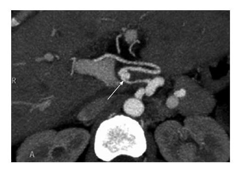
Figure 1 Contrast-enhanced-multidetector-row computed tomography-scan showing hepatic artery thrombosis after an endovascular intervention with stent placement.
Thrombus (arrow).
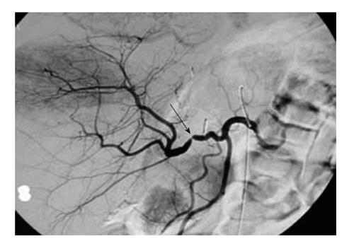
Figure 2 Arteriography showing an anastomotic hepatic artery stenosis after orthotopic liver transplantation.
Stenosis (arrow).
Figure 3 Contrast-enhanced-multidetector-row computed tomography-scan showing a hepatic artery pseudoaneurysm following orthotopic liver transplantation.
Pseudoaneurysm (arrow).
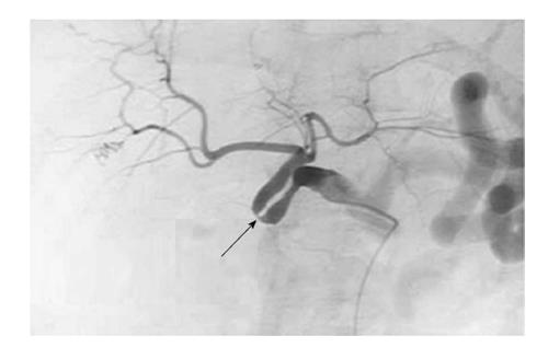
Figure 4 Arteriography showing a hepatic artery stenosis due to a kinking following orthotopic liver transplantation.
Kinking stenosis (arrow).
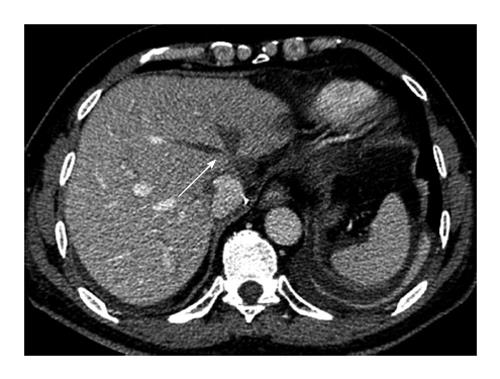
Figure 5 Contrast-enhanced-multidetector-row computed tomography-scan showing median and left thromboses hepatic veins following orthotopic liver transplantation (arrow).
- Citation: Piardi T, Lhuaire M, Bruno O, Memeo R, Pessaux P, Kianmanesh R, Sommacale D. Vascular complications following liver transplantation: A literature review of advances in 2015. World J Hepatol 2016; 8(1): 36-57
- URL: https://www.wjgnet.com/1948-5182/full/v8/i1/36.htm
- DOI: https://dx.doi.org/10.4254/wjh.v8.i1.36


