Copyright
©The Author(s) 2015.
World J Hepatol. Oct 28, 2015; 7(24): 2559-2562
Published online Oct 28, 2015. doi: 10.4254/wjh.v7.i24.2559
Published online Oct 28, 2015. doi: 10.4254/wjh.v7.i24.2559
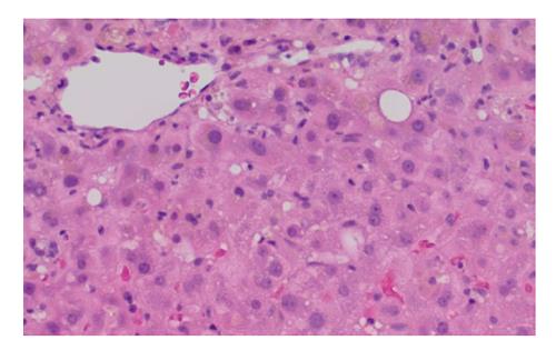
Figure 1 Histologic micrograph of liver biopsy.
This H and E photomicrograph shows a representative area of the adequate core needle biopsy, depicting overall a very mild lobular lymphocytic hepatitis with areas of mild cholestasis with canalicular plugs and focal hepatocyte bile pigmentation. The hepatic architecture is preserved with a normal relationship of central veins and portal tracts and no evidence of fibrosis.
- Citation: Ip S, Jeong R, Schaeffer DF, Yoshida EM. Unusual case of drug-induced cholestasis due to glucosamine and chondroitin sulfate. World J Hepatol 2015; 7(24): 2559-2562
- URL: https://www.wjgnet.com/1948-5182/full/v7/i24/2559.htm
- DOI: https://dx.doi.org/10.4254/wjh.v7.i24.2559


