Copyright
©The Author(s) 2015.
World J Hepatol. Oct 18, 2015; 7(23): 2432-2448
Published online Oct 18, 2015. doi: 10.4254/wjh.v7.i23.2432
Published online Oct 18, 2015. doi: 10.4254/wjh.v7.i23.2432
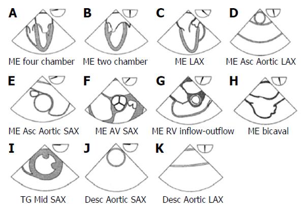
Figure 1 List of the 11 views suggested by the American Society of Echocardiography and Society of Cardiovascular Anesthesiologists guidelines on basic perioperative transoesophageal echocardiography.
Modified from Reeves et al[28]. ME: Mid esophageal; LAX: Long axis view.
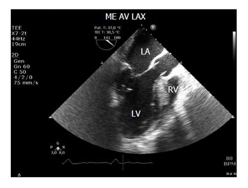
Figure 2 Transoesophageal echocardiography view (mid esophageal long axis view) used to measure left ventricular outflow tract diameter usually best imaged at a multiplane angle of 110°-140°.
AV: Aortic valve; LA: Left atrium; LV: Left ventricle; RV: Right ventricle; ME AV LAX: Mid esophageal aortic valve long axis view. Modified by Møller-Sørensen et al[45].
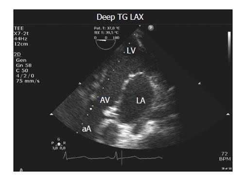
Figure 3 Transoesophageal echocardiography view (deep transgastric long axis view), used to measure velocity time integral.
AV: Aortic valve; LA: Left atrium; LV: Left ventricle; aA: Ascending aorta; TG LAX: Transgastric long axis view. Modified by Møller-Sørensen et al[45].
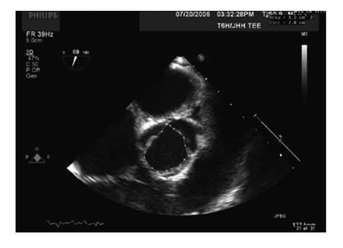
Figure 4 Transesophageal measurement of aortic valve planimetry from mid esophageal aortic valve SAX, usually best imaged at a multiplane angle of 40°-60°.
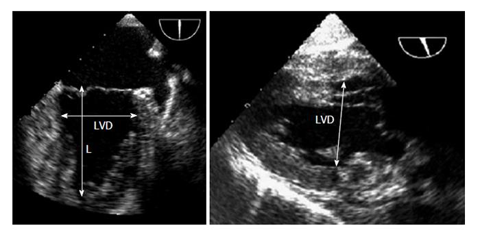
Figure 5 Transesophageal measurements of left ventricular length and minor left ventricular diameter from the ME 2C, usually best imaged at a multiplane angle of approximately 60°-90° and from the trans-gastric two-chamber view of the left ventricle, usually best imaged at an angle of approximately 90°-110°.
L: Length; LVD: Left ventricular diameter.
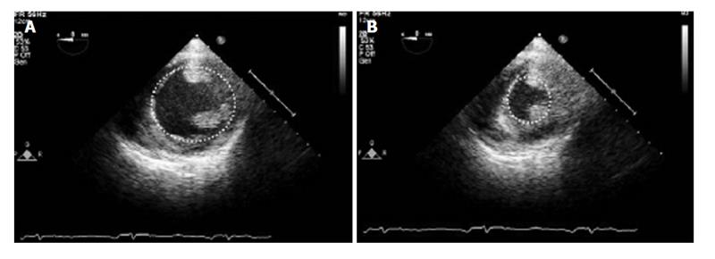
Figure 6 Transesophageal measurements of fractional area change.
Transgastric Mid SAX view of the left ventricle showing measurement of left ventricular end diastolic area (A) and left ventricular end-systolic area (B). The bidimensional image is usually best imaged at a multiplane angle of 0°. Modified by Guarracino Fabio et al[61].
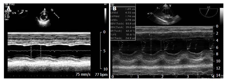
Figure 7 Transesophageal measurements of fractional shortening.
A: TG Mid SAX view of the left ventricle showing M-mode measurement of LVEDD and LVESD normalized for LVEDD. The bidimensional image is usually best imaged at a multiplane angle of 0°; B: TG LAX view of left ventricle showing M-Mode measurement of LVEDD and LVESD normalized for LVEDD usually best imaged at an angle of approximately 80°-110°. LAX: Long axis view; TG: Transgastric; LVEDD: Left ventricle end diastolic diameter; LVESD: Left ventricle end systolic diameter. Modified by Guarracino Fabio et al[61].
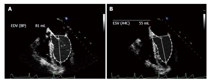
Figure 8 Calculation of ejection fraction using the disc method (Simpson's rule).
Transoesophageal echocardiography ME 4C view in diastole (A) and systole (B). The bidimensional image is usually best imaged at a multiplane angle of 0°. Modified by Guarracino Fabio et al[61]. EDV: End diastolic volume; ESV: End-systolic volume.
- Citation: De Pietri L, Mocchegiani F, Leuzzi C, Montalti R, Vivarelli M, Agnoletti V. Transoesophageal echocardiography during liver transplantation. World J Hepatol 2015; 7(23): 2432-2448
- URL: https://www.wjgnet.com/1948-5182/full/v7/i23/2432.htm
- DOI: https://dx.doi.org/10.4254/wjh.v7.i23.2432


