Copyright
©The Author(s) 2015.
World J Hepatol. Aug 28, 2015; 7(18): 2162-2170
Published online Aug 28, 2015. doi: 10.4254/wjh.v7.i18.2162
Published online Aug 28, 2015. doi: 10.4254/wjh.v7.i18.2162
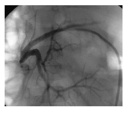
Figure 1 An 18-mo-old girl presented with a severe bilioenteric anastomotic stricture five months after receiving a left lateral segment graft from a live-donor.
After two percutaneous biliary interventions failed attempts to cross the stricture the patient underwent surgical revision and a new bilioenteric anastomosis was fashioned.
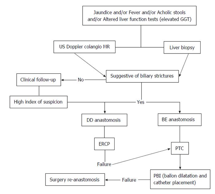
Figure 2 Proposed diagnostic and treatment algorithm for biliary strictures.
MR: Magnetic resonance; BE: Bilioenteric; PBI: Percutaneous biliary interventions; PTC: Percutaneous transhepatic cholangiography; DD: Duct-to-duct; US: Ultrasonography; GGT: Gamma-glutamyl transferase; ERCP: Endoscopic retrograde cholangio pancreatography.
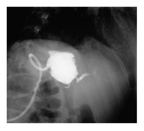
Figure 3 A 3-year-old boy developed a biloma 12-mo after receiving left lateral segment from a live-donor.
He had a persistent bile leak and segmental bile duct dilatation on abdominal computed tomography scan imaging. A fistulography was performed by injecting contrast through the pigtail catheter, and an excluded bile duct was evidenced.
- Citation: Feier FH, da Fonseca EA, Seda-Neto J, Chapchap P. Biliary complications after pediatric liver transplantation: Risk factors, diagnosis and management. World J Hepatol 2015; 7(18): 2162-2170
- URL: https://www.wjgnet.com/1948-5182/full/v7/i18/2162.htm
- DOI: https://dx.doi.org/10.4254/wjh.v7.i18.2162


