Copyright
©The Author(s) 2015.
World J Hepatol. Jun 18, 2015; 7(11): 1460-1483
Published online Jun 18, 2015. doi: 10.4254/wjh.v7.i11.1460
Published online Jun 18, 2015. doi: 10.4254/wjh.v7.i11.1460
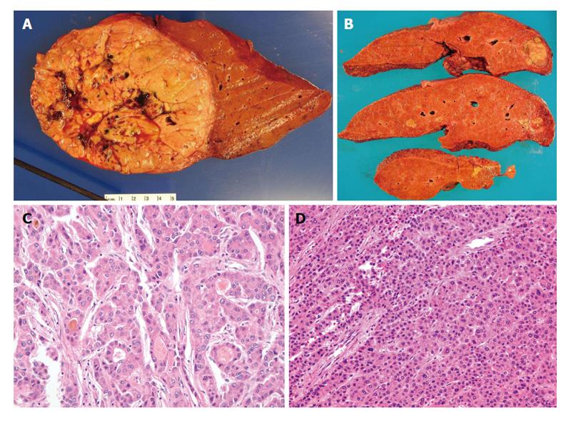
Figure 1 Pathology of classical hepatocellular carcinoma.
A: Gross photo of a well circumscribed, soft, yellowish to tan, and lobulated hepatocellular carcinoma (HCC) in a background of non-cirrhotic liver; B: Gross photo of a yellow and greenish, soft and lobulated HCC in a background of cirrhotic liver; C: Microphotos of HCC showing the pseudoacinar and pseudoglandular patterns, some containg the yellowish bile within the pseudoglandular structure with increased nuclear sizes; D: Microphotos of HCC showing thickened trabeculi, with increased unpaired arteries. Notice there are no normal structures present, i.e., portal tracts.
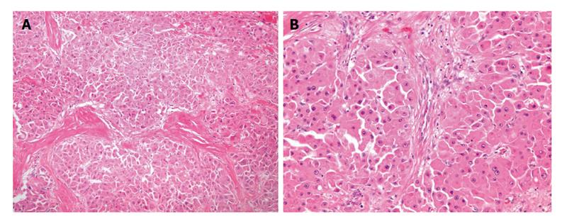
Figure 2 Pathology of fibrolamellar carcinoma.
A: Microphotos of fibrolamellar carcinoma showing the thick lamellar bands of fibrosis under low power magnification; B: In higher power magnification, the tumor cells are large and polygonal with abundant eosinophilic and granular cytoplasm, large vesiculated nuclei, and prominent nucleoli.
Figure 3 A 68-year-old male with cirrhosis and surgically proven hepatocellular carcinoma.
A: Thirty-four seconds after intravenous injection of ultrasound contrast (microbubbles) there is tumor (T with dashed line) enhancement; B: One and half minutes after injection the tumor (T with dashed line) is washing out of contrast. The images on the right side are a conventional sonogram (non-contrasted) of the lesion. The image on the left is a pulse inversion harmonics ultrasound for better visualization of ultrasound contrast media.
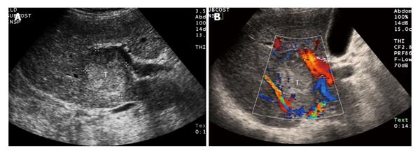
Figure 4 Ultrasound of hepatocellular carcinoma.
A: Ultrasound of the liver demonstrates a heterogeneous tumor (T) in the right lobe of the liver that was later characterized as definite hepatocellular carcinoma by computed tomography; B: Same lesion (T) using color Doppler ultrasound images to demonstrate blood flow in the adjacent vessels.
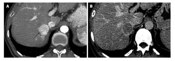
Figure 5 Computed tomography of hepatocellular carcinoma in 48-year-old male with hepatitis C.
A: Arterial phase contrast enhanced CT of the liver shows a strongly enhancing mass (arrow) in the right lobe, adjacent to the IVC. B: The same lesion (arrow) washes-out of contrast on the delayed phase and shows a thin capsule, this is diagnostic for HCC and corresponds to LI-RADS category 5. HCC: Hepatocellular carcinoma; CT: Computed tomography; LI-RADS: Liver imaging reporting and data system; IVC: Inferior vena cava.
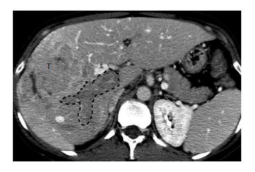
Figure 6 Portal vein invasion by hepatocellular carcinoma.
Computed tomography in portal venous phase shows a right lobe mass (T) and lack of enhancement of the portal vein (outlined), consistent with tumor invasion.
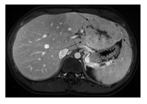
Figure 7 Magnetic resonance imaging of hepatocellular carcinoma in 19-year-old female.
Post contrast liver magnetic resonance imaging in portal venous phase shows a large mass (arrows) arising from the left lobe of a liver without cirrhosis. This lesion that has some imaging similarities with focal nodular hyperplasia, corresponded to fibrolamellar carcinoma on pathologic analysis.
Figure 8 Radiofrequency ablation of a focal hepatocellular carcinoma.
A: Contrast-enhanced MR of a 60-year-old male with cirrhosis demonstrates a single hepatocellular carcinoma in the right hepatic lobe (arrow); B: Ultrasound demonstrates a radiofrequency probe coursing through the hypoechoic tumor; C: Contrast-enhanced MR 1 mo after radio-frequency ablation demonstrates a large ablation defect, without any residual enhancement to suggest viable tumor. MR: Magnetic resonance.
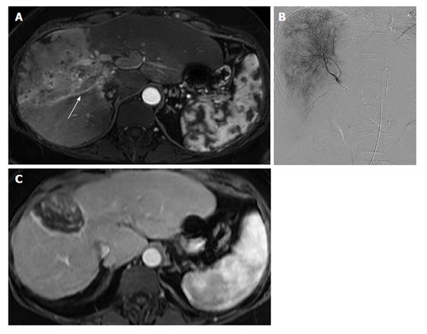
Figure 9 Yttrium-90 radioembolization of diffuse, infiltrative hepatocellular carcinoma with vascular invasion.
A: Contrast-enhanced MR of a 55-year-old female with cirrhosis demonstrates an infiltrative hepatocellular carcinoma replacing the anterior right hepatic lobe. Tumor-associated portal venous thrombus is present in the right portal vein (arrow); B: Digital subtraction angiogram with a microcatheter in the right hepatic artery shows diffuse tumor hypervascularity. The patient underwent a right hepatic artery Y90 radioembolization; C: Contrast-enhanced MR 12 mo after radioembolization demonstrates complete necrosis of the entire tumor with marked reduction in size. MR: Magnetic resonance; Y90: Yttrium-90.
- Citation: Yeh MM, Yeung RS, Apisarnthanarax S, Bhattacharya R, Cuevas C, Harris WP, Hon TLK, Padia SA, Park JO, Riggle KM, Daoud SS. Multidisciplinary perspective of hepatocellular carcinoma: A Pacific Northwest experience. World J Hepatol 2015; 7(11): 1460-1483
- URL: https://www.wjgnet.com/1948-5182/full/v7/i11/1460.htm
- DOI: https://dx.doi.org/10.4254/wjh.v7.i11.1460


