Copyright
©The Author(s) 2015.
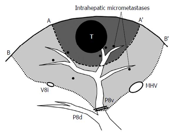
Figure 1 Concept of anatomic resection of the liver.
This schema shows an example of resection for hepatocellular carcinoma located in ventral part of Segment VIII. The line A-A’ indicates non-anatomic limited resection with adequate surgical margin and the line B-B’ represents anatomic resection of ventral part of Segment VIII. Because the “tumor-bearing” portal territory is at high risk of harboring micrometastases scattered via portal veins, systematic removal of the corresponding portal region would offer higher chance of eradicating cancer cells. T: Tumor; P8v: Ventral branch of Segment VIII portal pedicle; P8d: Dorsal branch of Segment VIII portal pedicle; MHV: Middle hepatic vein; V8i: Intermediate vein for Segment VIII.
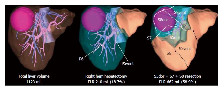
Figure 2 Surgical planning by three-dimensional simulation technique.
The three-dimensional liver simulation enables virtual hepatectomy and volume estimation of future liver remnant (FLR). In this case, a large hepatocellular carcinoma is located in segment 7 and 8, and partially extending to dorsal part of segment 5. Right hemihepatectomy is impossible due to very small FLR volume. However, when preserving the territory fed by a thick ventral branch for segment 5 (P5vent) and segment 6, the tumor is resectable leaving sufficient volume of the liver parenchyma. S5vent: Ventral part of segment 5; S5dor: Dorsal part of segment 5; S8vent: Ventral part of segment 8; S8dor: Dorsal part of segment 8.
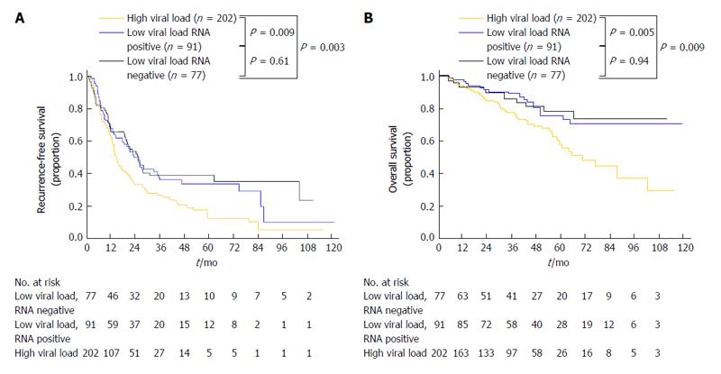
Figure 3 Cumulative recurrence rate (A) and cumulative overall survival (B) curves of low and high viral load groups stratified according to the results of hepatitis C virus RNA quantification.
(Adapted from Shindoh et al[60] with permission).
- Citation: Shindoh J, Hashimoto M, Watanabe G. Surgical approach for hepatitis C virus-related hepatocellular carcinoma. World J Hepatol 2015; 7(1): 70-77
- URL: https://www.wjgnet.com/1948-5182/full/v7/i1/70.htm
- DOI: https://dx.doi.org/10.4254/wjh.v7.i1.70


