Copyright
©2014 Baishideng Publishing Group Inc.
World J Hepatol. Jul 27, 2014; 6(7): 486-495
Published online Jul 27, 2014. doi: 10.4254/wjh.v6.i7.486
Published online Jul 27, 2014. doi: 10.4254/wjh.v6.i7.486
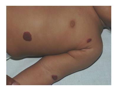
Figure 1 Cutaneous hemangiomatosis.
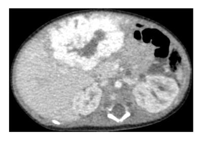
Figure 2 Abdominal computed tomography-contrast.
Focal hepatic hemangioma that shows centripetal enhancement and central sparing because of thrombosis, necrosis and/or intralesional hemorrhage.
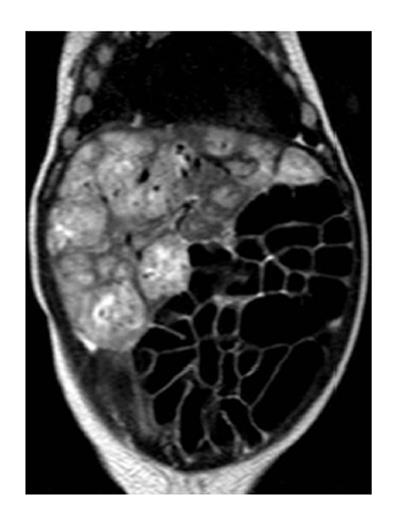
Figure 3 Abdominal magnetic resonance imaging-contrast.
Diffuse hepatic hemangioma that nearly totally replaces the liver.
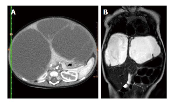
Figure 4 Abdominal computed tomography and magnetic resonance imaging.
A: Abdominal computed tomography-contrast shows enhancement of the solid component, septate, and the peripheral rim; B: Abdominal magnetic resonance imaging-contrast shows a high signal intensity on T2-weighted magnetic resonance sequences.
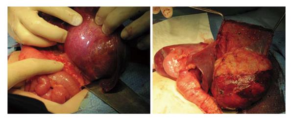
Figure 5 Surgical resection of mesenchymal hamartoma of the liver.
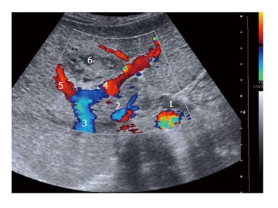
Figure 6 Doppler-US of the liver allows to investigate the relation between the tumor and the hepatic vessels.
1: Aorta; 2: Inferior vena cava; 3: Hepatic portal vein; 4: Left portal vein; 5: Right portal vein; 6 Tumor.
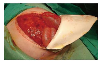
Figure 7 Extensive hepatic infiltration by neuroblastoma.
Surgical abdominal decompression by patch placement.
- Citation: Fernandez-Pineda I, Cabello-Laureano R. Differential diagnosis and management of liver tumors in infants. World J Hepatol 2014; 6(7): 486-495
- URL: https://www.wjgnet.com/1948-5182/full/v6/i7/486.htm
- DOI: https://dx.doi.org/10.4254/wjh.v6.i7.486


