Copyright
©2014 Baishideng Publishing Group Inc.
World J Hepatol. Jun 27, 2014; 6(6): 394-409
Published online Jun 27, 2014. doi: 10.4254/wjh.v6.i6.394
Published online Jun 27, 2014. doi: 10.4254/wjh.v6.i6.394
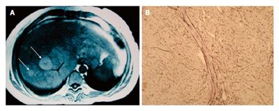
Figure 1 Regenerative nodular hyperplasia.
A: Magnetic resonance image of RNH (axial T1 FSE). Note the two hyperintense, solid nodules localized in the right hepatic lobe (arrows); B: Typical findings of a RNH lesion revealed by reticulin staining to highlight the sinusoidal architecture of the liver. Note the liver sinusoidal shrinking, mimicking a pseudonodule. RNH: Regenerative nodular hyperplasia.
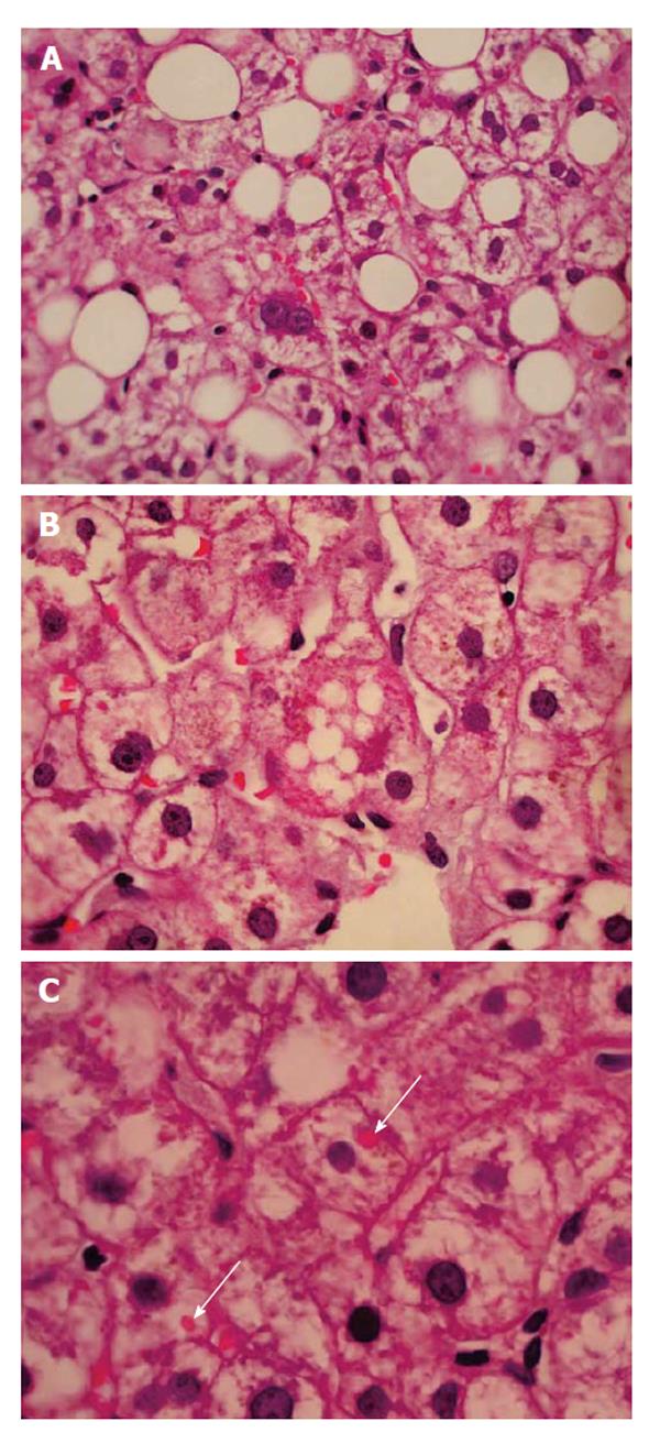
Figure 2 (A) Macrosteatosis, (B) microsteatosis and (C) megamitochondria (arrows), in a non-alcoholic 27-year-old patient with active systemic lupus erythematosus, treated with steroids and methotrexate (H and E staining).
- Citation: Bessone F, Poles N, Roma MG. Challenge of liver disease in systemic lupus erythematosus: Clues for diagnosis and hints for pathogenesis. World J Hepatol 2014; 6(6): 394-409
- URL: https://www.wjgnet.com/1948-5182/full/v6/i6/394.htm
- DOI: https://dx.doi.org/10.4254/wjh.v6.i6.394


