Copyright
©2013 Baishideng Publishing Group Co.
World J Hepatol. Jul 27, 2013; 5(7): 404-408
Published online Jul 27, 2013. doi: 10.4254/wjh.v5.i7.404
Published online Jul 27, 2013. doi: 10.4254/wjh.v5.i7.404
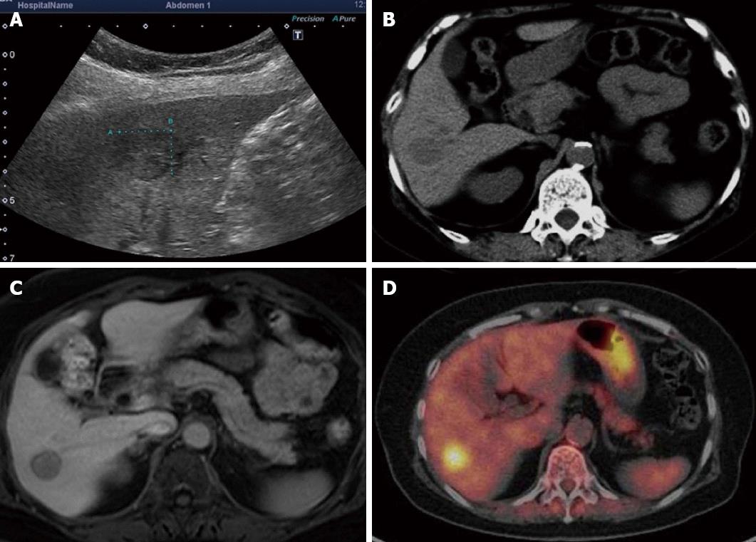
Figure 1 A mass approximately 15 mm in diameter was noted in the hepatic S6.
A: Ultrasonography; B: Computed tomography scan; C: Magnetic resonance imaging; D: Positron emission tomography.
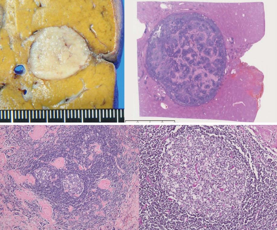
Figure 2 Lymph follicle hyperplasia was noted in the affected liver tissue.
Some follicles showed signs of vascular invasion, hyperplasia of the mantle layer, and the presence of multiple germ centers. Hyalinized interstitium was seen between follicles.
- Citation: Miyoshi H, Mimura S, Nomura T, Tani J, Morishita A, Kobara H, Mori H, Yoneyama H, Deguchi A, Himoto T, Yamamoto N, Okano K, Suzuki Y, Masaki T. A rare case of hyaline-type Castleman disease in the liver. World J Hepatol 2013; 5(7): 404-408
- URL: https://www.wjgnet.com/1948-5182/full/v5/i7/404.htm
- DOI: https://dx.doi.org/10.4254/wjh.v5.i7.404


