Copyright
©2013 Baishideng Publishing Group Co.
World J Hepatol. Jul 27, 2013; 5(7): 398-403
Published online Jul 27, 2013. doi: 10.4254/wjh.v5.i7.398
Published online Jul 27, 2013. doi: 10.4254/wjh.v5.i7.398
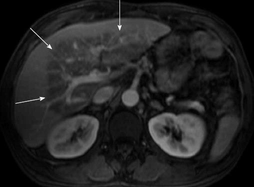
Figure 1 Spoiled gradient recalled-echo and fat-suppressed T1-weighted magnetic resonance imaging after iv gadolinium administration.
The venous phase on the axial portal plane shows, at the level of the left hepatic lobe and of the liver hilum region, a triangular hypo intense area (arrows) with hyper intense small areas within; vascular structures are preserved (May 2007).
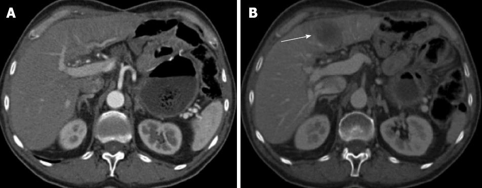
Figure 2 Axial computed tomography scan after iv iodinated contrast medium administration in the arterial hepatic phase (A) and portal phase (B).
A: The computed tomography study does not show focal lesions in the liver parenchyma (Jul 2008); B: A focal nodular hypo attenuated lesion is present affecting the left hepatic lobe (arrow); it presents with regular margins and appears mildly vascularised (Nov 2008).
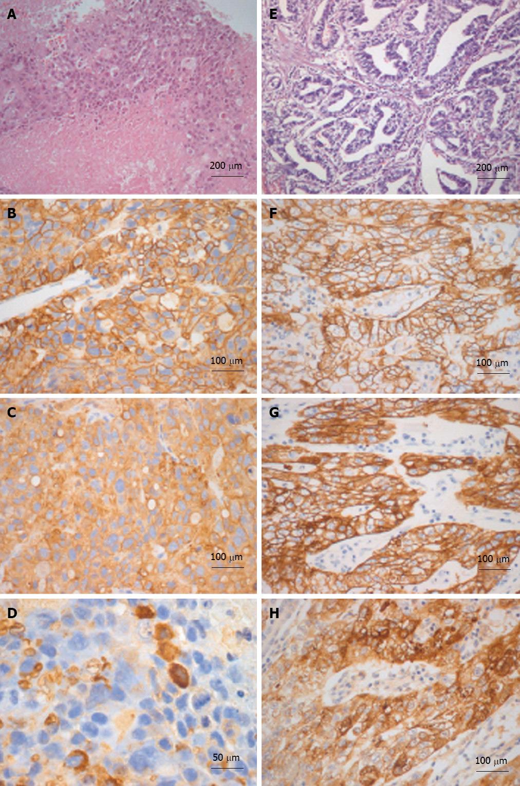
Figure 3 Histological-immunohistochemical characterizations of liver and gastric tumors are illustrated in A-D and E-H respectively.
The liver tumor is extensively necrotic (A) and neoplastic cells are immunoreactive for MOC-31 (B) and cytokeratin (CK)-18 (C). Sparse cells are immunoreactive for alpha-fetoprotein (AFP) as well (D). The gastric adenocarcinoma (E) is immunoreactive for MOC-31 (F), CK-18 (G) and AFP (H) as well. A and E: Hematoxylin and eosin.
- Citation: Cardinale V, De Filippis G, Corsi A, La Penna A, Rossi M, Catalano C, Bianco P, De Santis A, Alvaro D. An isolate alpha-fetoprotein producing gastric cancer liver metastasis emerged in a patient previously affected by radiation induced liver disease. World J Hepatol 2013; 5(7): 398-403
- URL: https://www.wjgnet.com/1948-5182/full/v5/i7/398.htm
- DOI: https://dx.doi.org/10.4254/wjh.v5.i7.398


