Copyright
©2013 Baishideng Publishing Group Co.
World J Hepatol. Jul 27, 2013; 5(7): 372-378
Published online Jul 27, 2013. doi: 10.4254/wjh.v5.i7.372
Published online Jul 27, 2013. doi: 10.4254/wjh.v5.i7.372
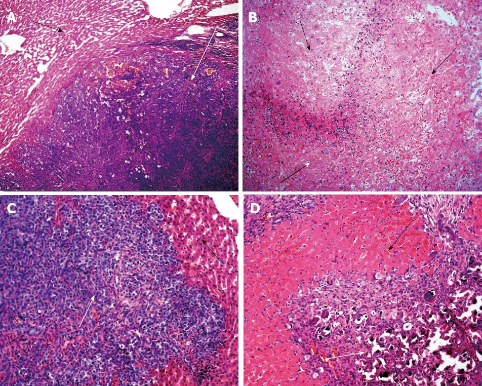
Figure 1 Photomicrograph of surgical specimen from liver VX2 tumor.
A: 24 h after saline injection revealing a large tumor nodule (white arrow) surrounded by normal hepatic tissue with no signs of necrosis (black arrow). (Hematoxylin-eosin, original magnification × 100); B: 24 h after 10% aspirin solution injection showing extensive necrotic areas (black arrow). (Hematoxylin-eosin, original magnification × 200); C: 7 d after saline injection demonstrating a large viable tumor nodule (white arrow) with normal adjacent hepatic parenchyma (black arrow). (Hematoxylin-eosin, original magnification × 200); D: 7 d after 10% aspirin solution injection showing normal hepatic parenchyma (black arrow) surrounded by inflammatory infiltrate and fibrosis (white arrows). (Hematoxylin-eosin, original magnification × 100).
-
Citation: Saad-Hossne R, Teixeira FV, Denadai R.
In vivo assessment of intratumoral aspirin injection to treat hepatic tumors. World J Hepatol 2013; 5(7): 372-378 - URL: https://www.wjgnet.com/1948-5182/full/v5/i7/372.htm
- DOI: https://dx.doi.org/10.4254/wjh.v5.i7.372


