Copyright
©2013 Baishideng Publishing Group Co.
World J Hepatol. Jun 27, 2013; 5(6): 336-339
Published online Jun 27, 2013. doi: 10.4254/wjh.v5.i6.336
Published online Jun 27, 2013. doi: 10.4254/wjh.v5.i6.336
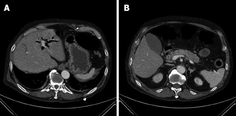
Figure 1 Computed tomography scans of the abdomen performed on admission.
A: Dilatation of intrahepatic bile ducts; B: Dilatation of extrahepatic bile ducts with wall thickening and contrast enhancement in the arterial phase of distal common bile duct and increased volume of the pancreas with loss of physiological lobular appearance.
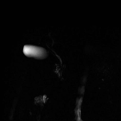
Figure 2 Magnetic resonance imaging, performed at the 12th week of follow up, showed a resolution of the radiological picture, with persistence of minimal dilation of the left biliary branch.
- Citation: Maida M, Macaluso FS, Cabibbo G, Lo Re G, Alessi N. Progressive multi-organ expression of immunoglobulin G4-related disease: A case report. World J Hepatol 2013; 5(6): 336-339
- URL: https://www.wjgnet.com/1948-5182/full/v5/i6/336.htm
- DOI: https://dx.doi.org/10.4254/wjh.v5.i6.336


