Copyright
©2013 Baishideng Publishing Group Co.
World J Hepatol. Apr 27, 2013; 5(4): 170-181
Published online Apr 27, 2013. doi: 10.4254/wjh.v5.i4.170
Published online Apr 27, 2013. doi: 10.4254/wjh.v5.i4.170
Figure 1 Hypothermia is associated with mortality in mice after 75% partial hepatectomy.
A: Survival outcome of mice that underwent either sham laparotomy (top solid line) or 75% partial hepatectomy (PH) (line with circles); B: Change in body temperature (BT) after 75% PH separated by survival outcome retrospectively. At 24 and 48 h after surgery, the BT of mice that finally died was significantly lower than that of mice that survived (P = 0.0001).
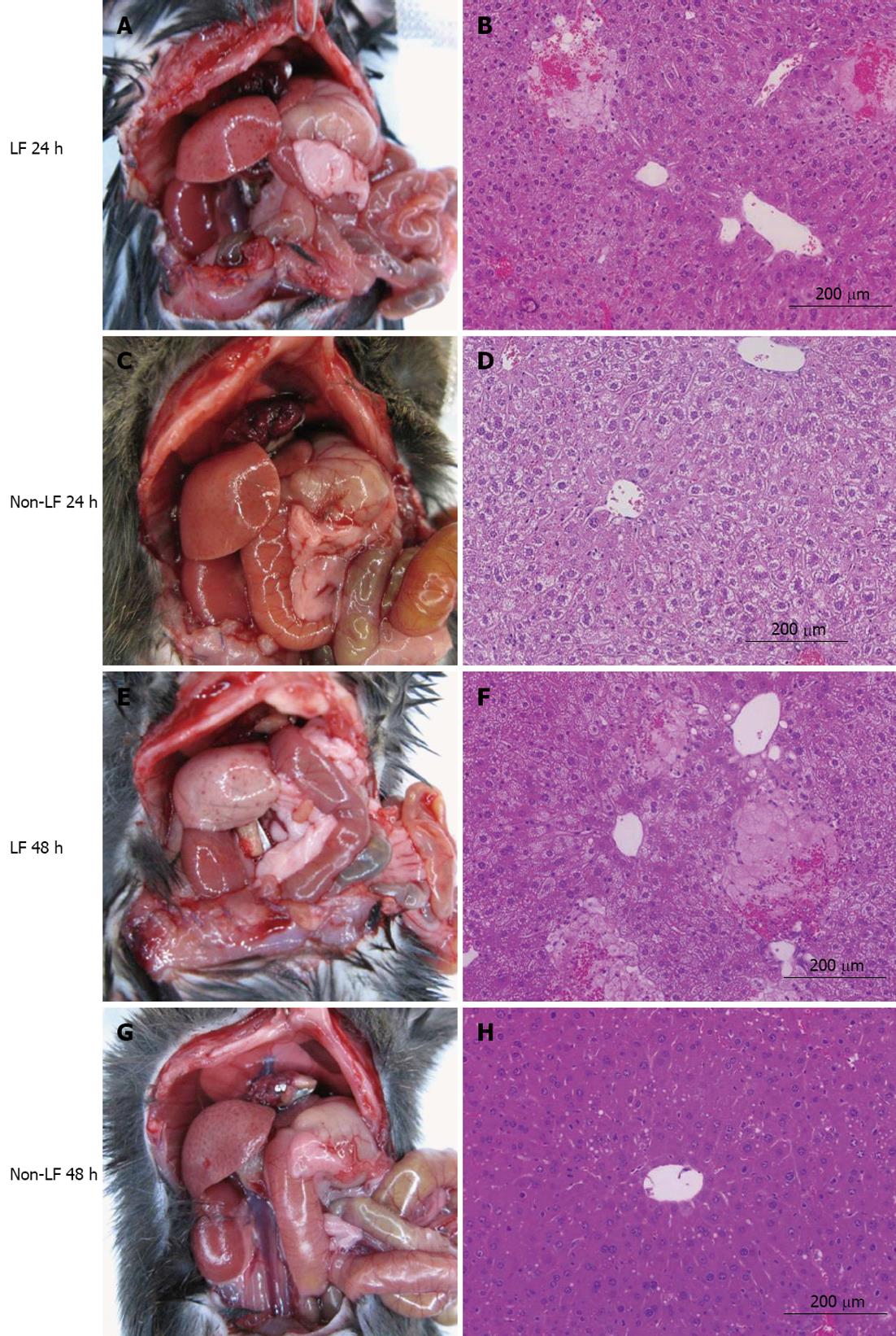
Figure 2 The gross appearance and hematoxylin-eosin-stained pathohistology of hypothermic and normothermic mice at 24 h (A-D) and 48 h (E-H) after 75% partial hepatectomy.
LF: Liver failure.
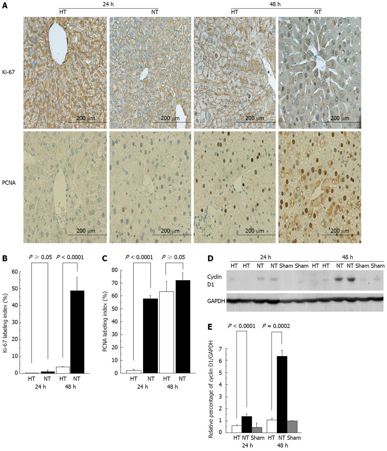
Figure 3 Hypothermic mice have a diminished proliferative index after 75% partial hepatectomy.
A: Ki-67 and proliferating cell nuclear antigen (PCNA) labeling at 24 and 48 h; B: Ki-67 labeling index; C: PCNA labeling index; D: Expression of cyclin D1; E: The ratio of cyclin D1/glyceraldehyde-3-phosphate dehydrogenase (GAPDH). HT: Hypothermic; NT: Normothermic.
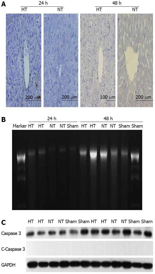
Figure 4 TdT-mediated DUTP-biotin nick end labeling, DNA laddering and caspase-3 assays to determine the contribution of apoptosis.
A: The TdT-mediated DUTP-biotin nick end labeling (TUNEL) immunostaining. TUNEL assay shows no differences between hypothermic (HT) and normothermic (NT) mice at 24 and 48 h; B: The DNA laddering. No DNA laddering patterns were detected at each time point; C: Caspase-3 expression. Cleaved (C) caspase-3 is not expressed. GAPDH: Glyceraldehyde-3-phosphate dehydrogenase.
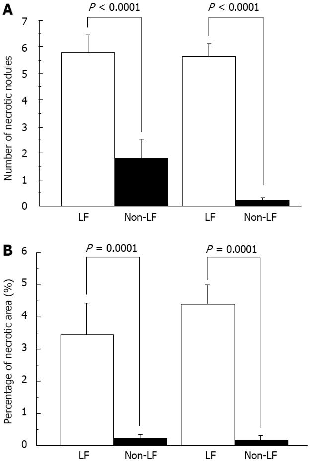
Figure 5 Necrosis occurs in the liver remnant of hypothermic mice after 75% partial hepatectomy.
A: Number of necrotic nodules in a ×100 microscope field in the liver remnant (LR); B: Percentage area of necrotic nodules in a LR section. LF: Liver failure.
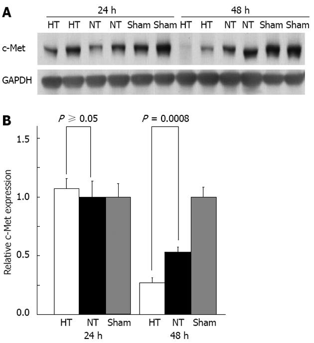
Figure 6 Signal transduction and energy production in the liver remnant after 75% partial hepatectomy.
A: Expression of c-Met; B: The expression of c-Met was the same in both study groups at 24 h but it was significantly lower in hypothermic mice compared with that in normothermic mice at 48 h (P = 0.0008). HT: Hypothermic; NT: Normothermic; GAPDH: Glyceraldehyde-3-phosphate dehydrogenase.
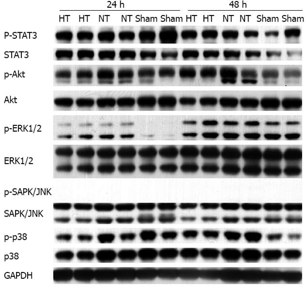
Figure 7 Actual findings of Western blot for p-STAT3, STAT3, p-AKT, AKT, p-ERK1/2,ERK1/2, p-SAPK/JNK, SAPK/JNK, p-p38, p38 and anti-glyceraldehyde-3-phosphate dehydrogenase.
Using Western blot, signal transduction in the liver remnant at 24 and 48 h in hypothermic (HT) and normothermic (NT) mice was examined, with sham animals serving as controls. Although p-STAT3, p-AKT, p-ERK1/2 and p-p38 were up-regulated in the liver remnant after 75% partial hepatectomy in both HT and NT mice compared with the sham controls, there was no significant difference between the HT and NT groups. There was no change in p-SAPK/JNK. Anti-glyceraldehyde-3-phosphate dehydrogenase (GAPDH) served as an internal control.
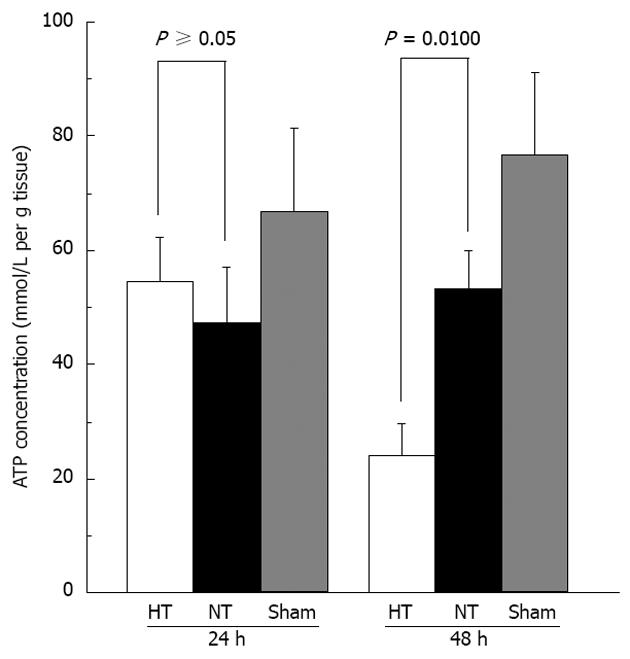
Figure 8 Adenosine triphosphate concentrations.
Adenosine triphosphate concentrations were determined in the liver remnant at 24 and 48 h in hypothermic and normothermic mice. Sham animals served as controls. HT: Hypothermic; NT: Normothermic.
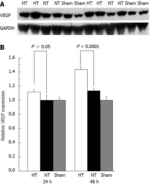
Figure 9 Vascular endothelial growth factor levels.
A: Expression of vascular endothelial growth factor (VEGF); B: Hypothermic mice had significantly higher VEGF levels than those in normothermic mice at 48 h but not at 24 h. HT: Hypothermic; NT: Normothermic; GAPDH: Glyceraldehyde-3-phosphate dehydrogenase.
- Citation: Ohashi N, Hori T, Uemoto S, Jermanus S, Chen F, Nakao A, Nguyen JH. Hypothermia predicts hepatic failure after extensive hepatectomy in mice. World J Hepatol 2013; 5(4): 170-181
- URL: https://www.wjgnet.com/1948-5182/full/v5/i4/170.htm
- DOI: https://dx.doi.org/10.4254/wjh.v5.i4.170


