Copyright
©2013 Baishideng Publishing Group Co.
World J Hepatol. Dec 27, 2013; 5(12): 696-700
Published online Dec 27, 2013. doi: 10.4254/wjh.v5.i12.696
Published online Dec 27, 2013. doi: 10.4254/wjh.v5.i12.696
Figure 1 Computer tomography scans of the abdomen performed on admission.
A: Arterial phase computer tomography (CT) scan shows small hepatocellular carcinoma nodule with mild enhancement (arrow) in right liver lobe; B: Hepatic venous phase; and C: Delayed phase CT scan at same level shows isoattenuation of lesion to liver parenchyma.
Figure 2 Computer tomography scans of the abdomen performed on admission.
A: Arterial phase computer tomography (CT) scan shows intense heterogeneous rim hyperenhancement of two round metastatic lesions in right liver lobe (arrows); B: Hepatic venous phase; and C: Delayed phase and CT scan show persistent rim enhancement around lesions (arrows). Diagnosis was confirmed at biopsy.
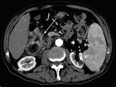
Figure 3 Computer tomography scans of the abdomen performed on admission.
Contrast material-enhanced axial computer tomography scan shows luminal narrowing and marked segmental circumferential thickening of the hepatic colon flexure. Adenocarcinoma was confirmed at colonoscopy and biopsy.
- Citation: Maida M, Macaluso FS, Galia M, Cabibbo G. Hepatocellular carcinoma and synchronous liver metastases from colorectal cancer in cirrhosis: A case report. World J Hepatol 2013; 5(12): 696-700
- URL: https://www.wjgnet.com/1948-5182/full/v5/i12/696.htm
- DOI: https://dx.doi.org/10.4254/wjh.v5.i12.696


