Copyright
©2013 Baishideng Publishing Group Co.
World J Hepatol. Nov 27, 2013; 5(11): 649-653
Published online Nov 27, 2013. doi: 10.4254/wjh.v5.i11.649
Published online Nov 27, 2013. doi: 10.4254/wjh.v5.i11.649
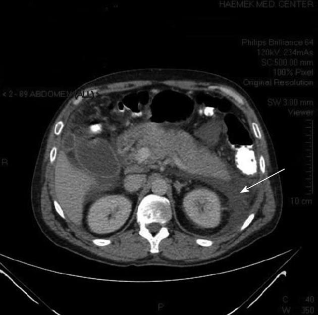
Figure 1 Abdominal computed tomography of the patient showing pancreatic edema and peripancreatic and pararenal fluid (arrow).
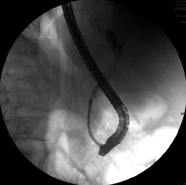
Figure 2 Endoscopic retrograde cholangiopancreatography of the patient showing removal of the stones by using a basket.
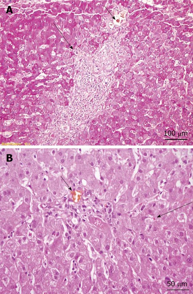
Figure 3 Liver biopsy.
A: Periodic acid Schiff-stained section (original magnification, × 200) showing mild interface hepatitis (long arrow) and a bile plug in the portal space bile duct (short arrow); B: Hematoxylin and eosin stain (original magnification, × 400) showing a bile plug in the portal space bile duct (short arrow) and mild intrahepatic cholestasis (long arrow).
- Citation: Nussinson E, Shahbari A, Shibli F, Chervinsky E, Trougouboff P, Markel A. Diagnostic challenges of Wilson’s disease presenting as acute pancreatitis, cholangitis, and jaundice. World J Hepatol 2013; 5(11): 649-653
- URL: https://www.wjgnet.com/1948-5182/full/v5/i11/649.htm
- DOI: https://dx.doi.org/10.4254/wjh.v5.i11.649


