Copyright
©2013 Baishideng Publishing Group Co.
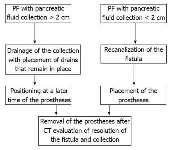
Figure 1 Flow chart of two-step (on the left) and one-step (on the right) procedures.
PF: Pancreatic fistula; CT: Computer tomography.
Figure 2 Diagnostic enhanced computer tomography scan (A), and un-enhanced computer tomography scan scans (B, C) performed during interventional procedure.
A: Patient developed a pancreatic fistula and abdominal fluid collection after surgery for pancreatic neoplasm; B, C: Percutaneous approach, was then adopted: images show trans-gastric puncture (B) and the access and placement of 2 drains in the collection(C).
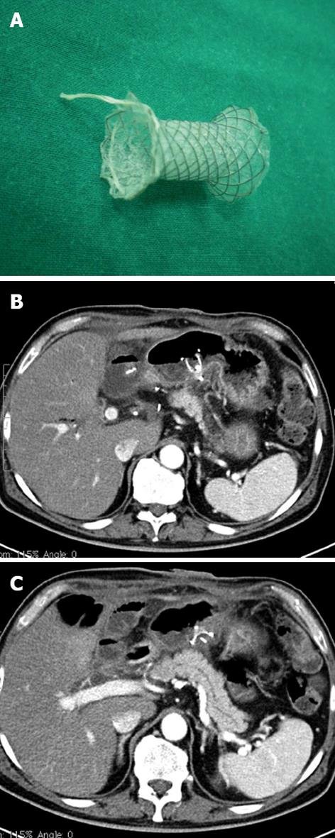
Figure 3 Prosthesis fully extended (Niti-S stent covered, TaeWoong Medical Co.
, Seoul, South Korea) (A) and enhanced computer tomography scans (B, C) performed after interventional procedure. Images demonstrate prosthesis in situ and resolution of peripancreatic collections.
Figure 4 The external fistula opens on the skin surface (A), a catheter has been positioned through the fistula (B) and two prostheses have then been placed through these two guides (C).
- Citation: Pradella S, Mazza E, Mondaini F, Colagrande S. Pancreatic fistula: A proposed percutaneous procedure. World J Hepatol 2013; 5(1): 33-37
- URL: https://www.wjgnet.com/1948-5182/full/v5/i1/33.htm
- DOI: https://dx.doi.org/10.4254/wjh.v5.i1.33


