Copyright
©2012 Baishideng.
World J Hepatol. Aug 27, 2012; 4(8): 252-255
Published online Aug 27, 2012. doi: 10.4254/wjh.v4.i8.252
Published online Aug 27, 2012. doi: 10.4254/wjh.v4.i8.252
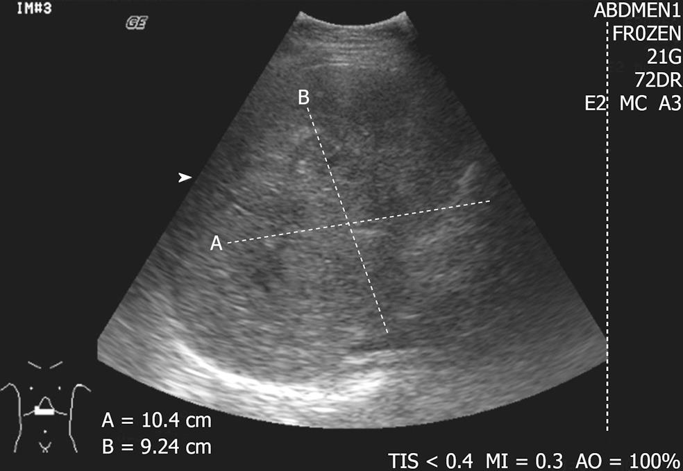
Figure 1 Ultrasound of liver showed mixed heterogeneous echogenicity lesions.
Ill defined internal hyperechogenicity with “dirty shadow” appearance suspicious of gas content.
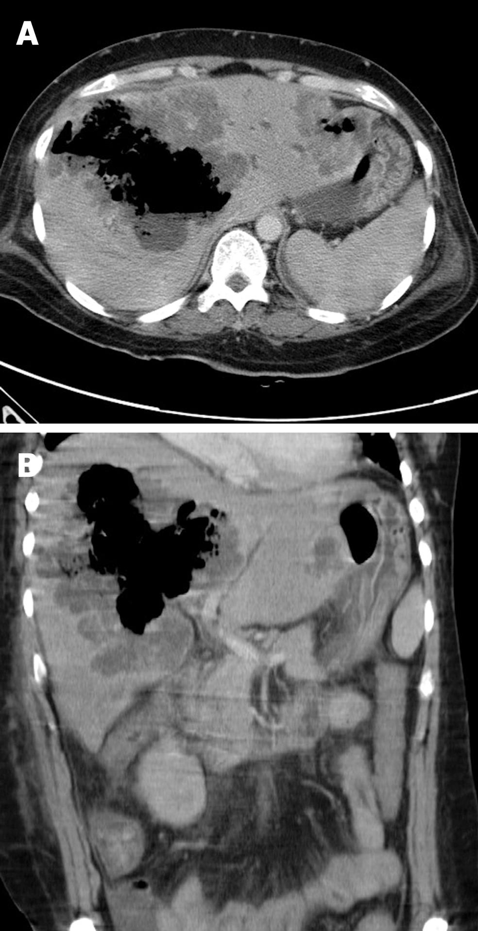
Figure 2 Axial (A) and sagittal (B) contrast multi-detector computerized tomography scan of abdomen.
Rim-enhancing cystic lesions with internal gas content occupying both hepatic lobes with the largest occupying the right lobe.
-
Citation: Law ST, Lee MK. A middle-aged lady with a pyogenic liver abscess caused by
Clostridium perfringens . World J Hepatol 2012; 4(8): 252-255 - URL: https://www.wjgnet.com/1948-5182/full/v4/i8/252.htm
- DOI: https://dx.doi.org/10.4254/wjh.v4.i8.252


