Copyright
©2012 Baishideng Publishing Group Co.
World J Hepatol. Dec 27, 2012; 4(12): 419-421
Published online Dec 27, 2012. doi: 10.4254/wjh.v4.i12.419
Published online Dec 27, 2012. doi: 10.4254/wjh.v4.i12.419
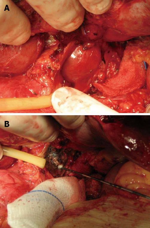
Figure 1 The exposure and radiofrequency ablation of the lesion in the caudate lobe under laparotomy.
A: The tumor found in the caudate lobe. The arrow indicates the lesion; B: The largest tumor in the Caudate lobe measuring 4 cm was treated with radiofrequency (RF) ablation, with RF electrodes were placed at the tumor site.
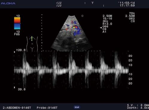
Figure 2 Introperative ultrosound study showed a hepatic artery pseudoaneurysm within the hematoma.
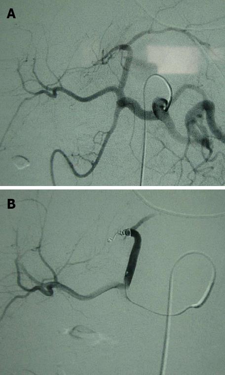
Figure 3 Hepatic angiography before and after coil embolization.
A: Hepatic angiography revealed a pseudoaneurysm in the lower left hepatic artery; B; Hepatic angiography immediately after coil embolization. Coils were placed using a 2.1 F microcatheter into and proximal to the pseudoaneurysm. The pseudoaneurysm was then excluded.
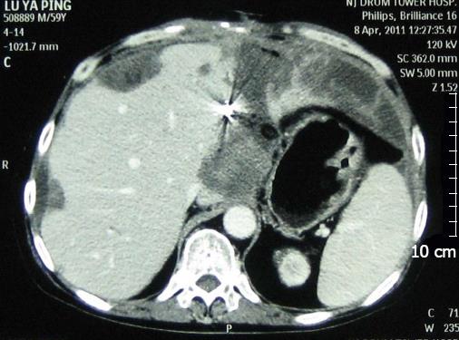
Figure 4 Computed tomography scan acquired 1 mo later.
The image revealed that the pseudoaneurysm had disappeared. No tumor enhancement was apparent at the ablated site. The coils used for embolization were visualized.
- Citation: Wu XY, Shi XL, Zhou JX, Qiu YD, Zhou T, Han B, Ding YT. Life-threatening hemorrhage after liver radiofrequency ablation successfully controlled by transarterial embolization. World J Hepatol 2012; 4(12): 419-421
- URL: https://www.wjgnet.com/1948-5182/full/v4/i12/419.htm
- DOI: https://dx.doi.org/10.4254/wjh.v4.i12.419


