Copyright
©2012 Baishideng Publishing Group Co.
World J Hepatol. Dec 27, 2012; 4(12): 394-398
Published online Dec 27, 2012. doi: 10.4254/wjh.v4.i12.394
Published online Dec 27, 2012. doi: 10.4254/wjh.v4.i12.394
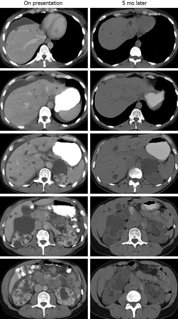
Figure 1 Contrast-enhanced abdominal computed tomography.
Left panel: Serial images from an axial computed tomography (CT) scan at the time of the patient’s presentation to the local emergency department, showing perihepatic ascites and multiple liver and kidney cysts; Right panel: Repeat abdominal CT scan five months following the acute presentation, showing corresponding images of multiple liver and kidney cysts without perihepatic ascites.
- Citation: Chaudhary S, Qian Q. Acute abdomen and ascites as presenting features of autosomal dominant polycystic kidney disease. World J Hepatol 2012; 4(12): 394-398
- URL: https://www.wjgnet.com/1948-5182/full/v4/i12/394.htm
- DOI: https://dx.doi.org/10.4254/wjh.v4.i12.394


