Copyright
©2010 Baishideng Publishing Group Co.
World J Hepatol. Aug 27, 2010; 2(8): 322-324
Published online Aug 27, 2010. doi: 10.4254/wjh.v2.i8.322
Published online Aug 27, 2010. doi: 10.4254/wjh.v2.i8.322
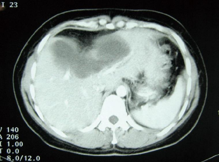
Figure 1 Computed tomography showing the hepatic cyst in segments III, IV and V with septations and intimate contact with portal veins.
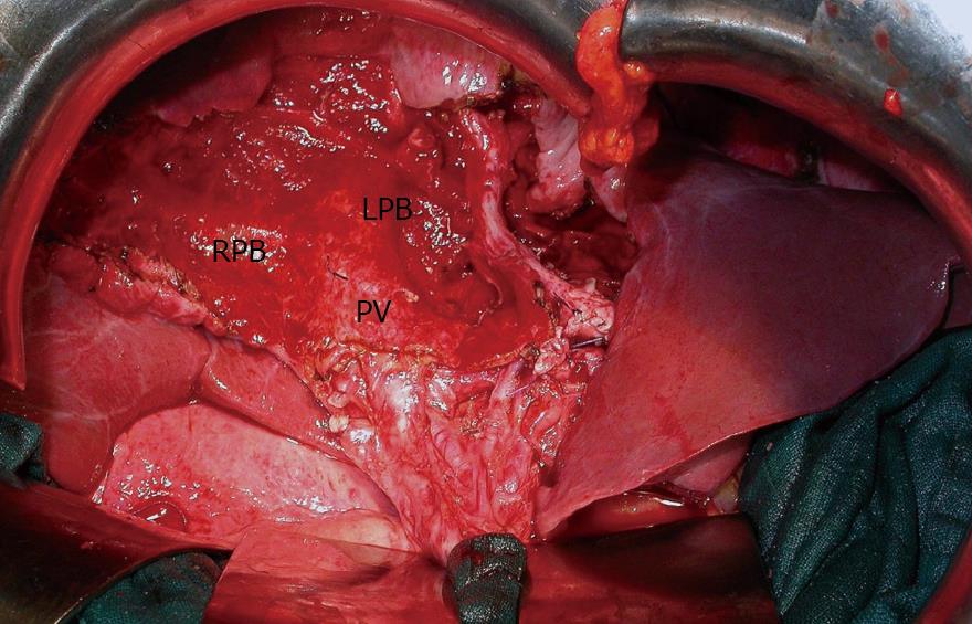
Figure 2 Operative photography after complete removal of the cyst: central hepatectomy.
PV: portal vein; RPB: right portal vein; LPB: left portal vein.
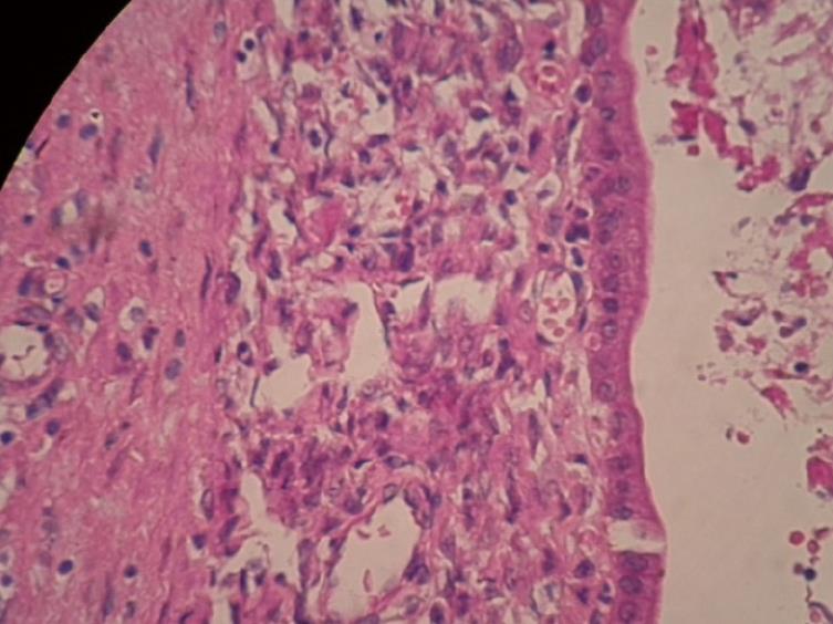
Figure 3 Photomicrograph demonstrating a cuboidal epithelial lining with dense spindle cell (mesenchymal) stroma.
- Citation: Elfadili H, Majbar A, Zouaidia F, Elamrani N, Sabbah F, Raiss M, Mahassini N, Hrora A, Ahallat M. Spontaneous rupture of a recurrent hepatic cystadenoma. World J Hepatol 2010; 2(8): 322-324
- URL: https://www.wjgnet.com/1948-5182/full/v2/i8/322.htm
- DOI: https://dx.doi.org/10.4254/wjh.v2.i8.322


